
- school Campus Bookshelves
- menu_book Bookshelves
- perm_media Learning Objects
- login Login
- how_to_reg Request Instructor Account
- hub Instructor Commons

Margin Size
- Download Page (PDF)
- Download Full Book (PDF)
- Periodic Table
- Physics Constants
- Scientific Calculator
- Reference & Cite
- Tools expand_more
- Readability
selected template will load here
This action is not available.
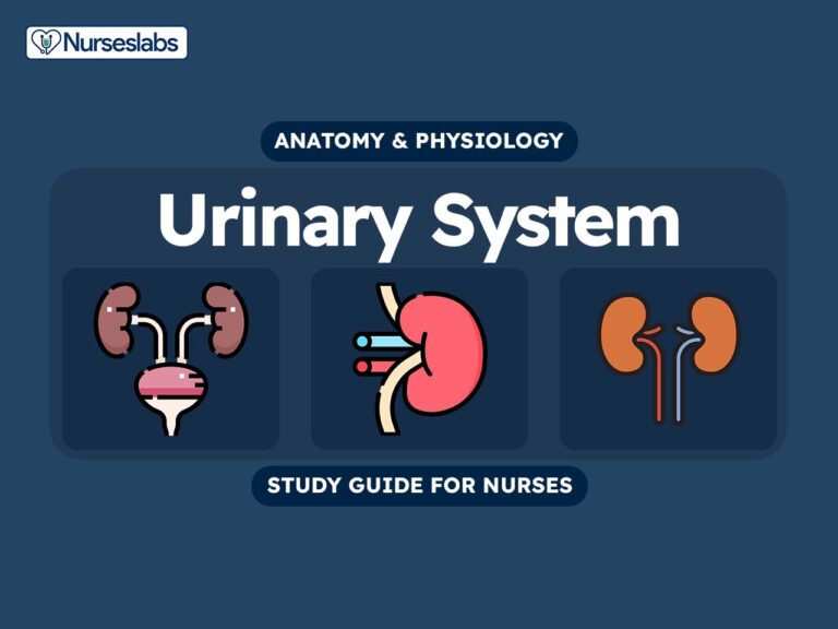
22.1: Introduction to the Urinary System
- Last updated
- Save as PDF
- Page ID 22417

- Whitney Menefee, Julie Jenks, Chiara Mazzasette, & Kim-Leiloni Nguyen
- Reedley College, Butte College, Pasadena City College, & Mt. San Antonio College via ASCCC Open Educational Resources Initiative
\( \newcommand{\vecs}[1]{\overset { \scriptstyle \rightharpoonup} {\mathbf{#1}} } \)
\( \newcommand{\vecd}[1]{\overset{-\!-\!\rightharpoonup}{\vphantom{a}\smash {#1}}} \)
\( \newcommand{\id}{\mathrm{id}}\) \( \newcommand{\Span}{\mathrm{span}}\)
( \newcommand{\kernel}{\mathrm{null}\,}\) \( \newcommand{\range}{\mathrm{range}\,}\)
\( \newcommand{\RealPart}{\mathrm{Re}}\) \( \newcommand{\ImaginaryPart}{\mathrm{Im}}\)
\( \newcommand{\Argument}{\mathrm{Arg}}\) \( \newcommand{\norm}[1]{\| #1 \|}\)
\( \newcommand{\inner}[2]{\langle #1, #2 \rangle}\)
\( \newcommand{\Span}{\mathrm{span}}\)
\( \newcommand{\id}{\mathrm{id}}\)
\( \newcommand{\kernel}{\mathrm{null}\,}\)
\( \newcommand{\range}{\mathrm{range}\,}\)
\( \newcommand{\RealPart}{\mathrm{Re}}\)
\( \newcommand{\ImaginaryPart}{\mathrm{Im}}\)
\( \newcommand{\Argument}{\mathrm{Arg}}\)
\( \newcommand{\norm}[1]{\| #1 \|}\)
\( \newcommand{\Span}{\mathrm{span}}\) \( \newcommand{\AA}{\unicode[.8,0]{x212B}}\)
\( \newcommand{\vectorA}[1]{\vec{#1}} % arrow\)
\( \newcommand{\vectorAt}[1]{\vec{\text{#1}}} % arrow\)
\( \newcommand{\vectorB}[1]{\overset { \scriptstyle \rightharpoonup} {\mathbf{#1}} } \)
\( \newcommand{\vectorC}[1]{\textbf{#1}} \)
\( \newcommand{\vectorD}[1]{\overrightarrow{#1}} \)
\( \newcommand{\vectorDt}[1]{\overrightarrow{\text{#1}}} \)
\( \newcommand{\vectE}[1]{\overset{-\!-\!\rightharpoonup}{\vphantom{a}\smash{\mathbf {#1}}}} \)
Chapter Learning Objectives
- Label structures of the urinary system
- Characterize the roles of each of the parts of the urinary system
- Illustrate the macroscopic and microscopic structures of the kidney
- Trace the flow of blood and urine through the kidney
- Trace the flow of urine through the urinary tract
This chapter will help you to understand the anatomy of the urinary system: kidneys, ureters, urinary bladder, and urethra. You will trace the blood flow through the kidney; think of the kidney as a regulator of plasma makeup rather than simply a urine producer. Knowing the anatomy will help you to understand how the urinary system maintains homeostasis and affects all the other systems of the body when you take physiology.
Interactive Link
Watch this video Kidney Function from the Howard Hughes Medical Institute for an introduction to the urinary system.
The Urinary or Renal System consists of two kidneys and the pathway, two ureters, urinary bladder, and one urethra, for urine to exit the body. The kidneys' primary function is to make urine by first filtering out many of the electrolytes, solutes, and water in the blood, forming the filtrate. As the filtrate passes through microscopic units called nephrons, certain solutes and water molecules are reabsorbed back to the blood because they are too precious to waste and other molecules are secreted because the body does not want them. As the filtrate becomes more concentrated, because water has been reabsorbed back to the blood, as it moves through the nephron, the end product is urine containing the waste products that the body can spare. Another perspective on the function of the kidneys is to monitor the level of certain solutes in the blood to maintain homeostasis. The kidneys' secondary function is to make hormones. Please review the Endocrine chapter for details.
The two ureters will carry urine from the kidneys down to the urinary bladder by peristalsis for storage. As the urinary bladder fills with urine, nerve endings will detect the stretched bladder to signal emptying. With sphincters relaxing, urine will flow out of the urinary bladder through the urethra to be expelled.
Contributors and Attributions
OpenStax Anatomy & Physiology (CC BY 4.0). Access for free at https://openstax.org/books/anatomy-and-physiology
- View All Systems
- Anatomy Systems
- Cardiovascular
- Respiratory
- Integumentary
- View All Telehealth
- Online Pharmacies
- Hims Review
- Keeps Review
- Keeps vs Hims
- BlueChew Review
- Rugiet Review
- Hims ED Review
- Online Therapy
- BetterHelp Review
- BetterHelp vs Talkspace
- View All Testing
- General Health Tests
- Best DNA Health Test
- Best Microbiome Test
- Viome Review
- Best At-Home Herpes Test
- STDcheck Review
- View All Self-Care
- Hair and Hair Loss
- Best Hair Loss Treatment for Men
- Best Laser Cap for Hair Loss
- Kiierr Laser Cap Review
- Nutrition Guide
- Found Weight Loss Review
- Relationships
- Best STD Dating Websites
- Best Herpes Dating Websites
- Sexual Health
- Best Erectile Dysfunction Treatment
- Best Delay Spray
- Best Prostate Massager
- BlueChew Free Trial
- Best CBD for Arthritis
- Best Incontinence Underwear for Women
- Best Incontinence Underwear for Men
- All Health Products
- Best Amino Acid Supplements
- Best Muscle Building Stack
- Best NMN Supplement
- Best NAC Supplement
- Best NAD Supplement
- Best Berberine Supplement
- Mood and Energy
- Best Nootropics
- Best Saffron Supplement
- Fitness and Weight
- Vital Reds Review
- Total Restore Review
- Best Testosterone Booster
- Best Male Enhancement Pills
- Best Volume Pills
- View All Research
- Skeletal System
- Muscular System
- Cardiovascular System
- Digestive System
- Nervous System
- Respiratory System
- Urinary System
The Urinary System
Explore the anatomy and physiology of the kidneys, ureters, urinary bladder, and urethra, learning all about how our bodies filter out and remove waste through urine..
- 2D Interactive
- NEW 3D Rotate and Zoom
Anatomy Explorer
- LOWER TORSO
Urinary Bladder
Change current view angle.
- Urinary System (Male View)
- Urinary System (Male Posterior View)
- Urinary System (Posterior View)
Toggle Anatomy System
- Endocrine System
- Female Reproductive System
- Immune and Lymphatic Systems
- Integumentary System
- Male Reproductive System
Switch Gender
Anatomy term.
Displayed on other page

The urinary system consists of the kidneys, ureters, urinary bladder, and urethra. The kidneys filter the blood to remove wastes and produce urine. The ureters, urinary bladder, and urethra together form the urinary tract, which acts as a plumbing system to drain urine from the kidneys, store it, and then release it during urination. Besides filtering and eliminating wastes from the body, the urinary system also maintains the homeostasis of water, ions, pH, blood pressure, calciummycontentbreak and red blood cells.
Urinary System Anatomy
The kidneys are a pair of bean-shaped organs found along the posterior wall of the abdominal cavity. The left kidney is located slightly higher than the right kidney because the right side of the liver is much larger than the left side. The kidneys, unlike the other organs of the abdominal cavity, are located posterior to the peritoneum and touch the muscles of the back . The kidneys are surrounded by a layer of adipose that holds them in place and protects them from physical damage. The kidneys filter metabolic wastes, excess ions, and chemicals from the blood to form urine.
The ureters are a pair of tubes that carry urine from the kidneys to the urinary bladder. The ureters are about 10 to 12 inches long and run on the left and right sides of the body parallel to the vertebral column . Gravity and peristalsis of smooth muscle tissue in the walls of the ureters move urine toward the urinary bladder. The ends of the ureters extend slightly into the urinary bladder and are sealed at the point of entry to the bladder by the ureterovesical valves. These valves prevent urine from flowing back towards the kidneys.
The urinary bladder is a sac-like hollow organ used for the storage of urine. The urinary bladder is located along the body’s midline at the inferior end of the pelvis . Urine entering the urinary bladder from the ureters slowly fills the hollow space of the bladder and stretches its elastic walls. The walls of the bladder allow it to stretch to hold anywhere from 600 to 800 milliliters of urine.
The urethra is the tube through which urine passes from the bladder to the exterior of the body. The female urethra is around 2 inches long and ends inferior to the clitoris and superior to the vaginal opening. In males, the urethra is around 8 to 10 inches long and ends at the tip of the penis . The urethra is also an organ of the male reproductive system as it carries sperm out of the body through the penis.

The flow of urine through the urethra is controlled by the internal and external urethral sphincter muscles. The internal urethral sphincter is made of smooth muscle and opens involuntarily when the bladder reaches a certain set level of distention. The opening of the internal sphincter results in the sensation of needing to urinate. The external urethral sphincter is made of skeletal muscle and may be opened to allow urine to pass through the urethra or may be held closed to delay urination.
Urinary System Physiology
Maintenance of homeostasis.
The kidneys maintain the homeostasis of several important internal conditions by controlling the excretion of substances out of the body.
The kidney can control the excretion of potassium, sodium, calcium, magnesium, phosphate, and chloride ions into urine. In cases where these ions reach a higher than normal concentration, the kidneys can increase their excretion out of the body to return them to a normal level. Conversely, the kidneys can conserve these ions when they are present in lower than normal levels by allowing the ions to be reabsorbed into the blood during filtration. (See more about ions .)
The kidneys monitor and regulate the levels of hydrogen ions (H+) and bicarbonate ions in the blood to control blood pH. H+ ions are produced as a natural byproduct of the metabolism of dietary proteins and accumulate in the blood over time. The kidneys excrete excess H+ ions into urine for elimination from the body. The kidneys also conserve bicarbonate ions, which act as important pH buffers in the blood.
The cells of the body need to grow in an isotonic environment in order to maintain their fluid and electrolyte balance. The kidneys maintain the body’s osmotic balance by controlling the amount of water that is filtered out of the blood and excreted into urine. When a person consumes a large amount of water, the kidneys reduce their reabsorption of water to allow the excess water to be excreted in urine. This results in the production of dilute, watery urine. In the case of the body being dehydrated, the kidneys reabsorb as much water as possible back into the blood to produce highly concentrated urine full of excreted ions and wastes. The changes in excretion of water are controlled by antidiuretic hormone (ADH). ADH is produced in the hypothalamus and released by the posterior pituitary gland to help the body retain water.
Blood Pressure
The kidneys monitor the body’s blood pressure to help maintain homeostasis. When blood pressure is elevated, the kidneys can help to reduce blood pressure by reducing the volume of blood in the body. The kidneys are able to reduce blood volume by reducing the reabsorption of water into the blood and producing watery, dilute urine. When blood pressure becomes too low, the kidneys can produce the enzyme renin to constrict blood vessels and produce concentrated urine, which allows more water to remain in the blood.
Inside each kidney are around a million tiny structures called nephrons. The nephron is the functional unit of the kidney that filters blood to produce urine. Arterioles in the kidneys deliver blood to a bundle of capillaries surrounded by a capsule called a glomerulus . As blood flows through the glomerulus, much of the blood’s plasma is pushed out of the capillaries and into the capsule, leaving the blood cells and a small amount of plasma to continue flowing through the capillaries. The liquid filtrate in the capsule flows through a series of tubules lined with filtering cells and surrounded by capillaries. The cells surrounding the tubules selectively absorb water and substances from the filtrate in the tubule and return it to the blood in the capillaries. At the same time, waste products present in the blood are secreted into the filtrate. By the end of this process, the filtrate in the tubule has become urine containing only water, waste products, and excess ions. The blood exiting the capillaries has reabsorbed all of the nutrients along with most of the water and ions that the body needs to function.
Storage and Excretion of Wastes
After urine has been produced by the kidneys, it is transported through the ureters to the urinary bladder. The urinary bladder fills with urine and stores it until the body is ready for its excretion. When the volume of the urinary bladder reaches anywhere from 150 to 400 milliliters, its walls begin to stretch and stretch receptors in its walls send signals to the brain and spinal cord . These signals result in the relaxation of the involuntary internal urethral sphincter and the sensation of needing to urinate. Urination may be delayed as long as the bladder does not exceed its maximum volume, but increasing nerve signals lead to greater discomfort and desire to urinate.
Urination is the process of releasing urine from the urinary bladder through the urethra and out of the body. The process of urination begins when the muscles of the urethral sphincters relax, allowing urine to pass through the urethra. At the same time that the sphincters relax, the smooth muscle in the walls of the urinary bladder contract to expel urine from the bladder.
Production of Hormones
The kidneys produce and interact with several hormones that are involved in the control of systems outside of the urinary system.
Calcitriol is the active form of vitamin D in the human body. It is produced by the kidneys from precursor molecules produced by UV radiation striking the skin. Calcitriol works together with parathyroid hormone (PTH) to raise the level of calcium ions in the bloodstream. When the level of calcium ions in the blood drops below a threshold level, the parathyroid glands release PTH, which in turn stimulates the kidneys to release calcitriol. Calcitriol promotes the small intestine to absorb calcium from food and deposit it into the bloodstream. It also stimulates the osteoclasts of the skeletal system to break down bone matrix to release calcium ions into the blood.
Erythropoietin
Erythropoietin, also known as EPO, is a hormone that is produced by the kidneys to stimulate the production of red blood cells. The kidneys monitor the condition of the blood that passes through their capillaries, including the oxygen-carrying capacity of the blood. When the blood becomes hypoxic, meaning that it is carrying deficient levels of oxygen, cells lining the capillaries begin producing EPO and release it into the bloodstream. EPO travels through the blood to the r ed bone marrow , where it stimulates hematopoietic cells to increase their rate of red blood cell production. Red blood cells contain hemoglobin, which greatly increases the blood’s oxygen-carrying capacity and effectively ends the hypoxic conditions.
Renin is not a hormone itself, but an enzyme that the kidneys produce to start the renin-angiotensin system (RAS). The RAS increases blood volume and blood pressure in response to low blood pressure, blood loss, or dehydration. Renin is released into the blood where it catalyzes angiotensinogen from the liver into angiotensin I. Angiotensin I is further catalyzed by another enzyme into Angiotensin II.
Angiotensin II stimulates several processes, including stimulating the adrenal cortex to produce the hormone aldosterone. Aldosterone then changes the function of the kidneys to increase the reabsorption of water and sodium ions into the blood, increasing blood volume and raising blood pressure. Negative feedback from increased blood pressure finally turns off the RAS to maintain healthy blood pressure levels.
Other Recommended Resources
- BetterHelp Reviews
- Fast HSDD Treatments for Women
- Quick Extender Pro Reviews
- Best Extender
- Best Vacuum Pump for Erectile Dysfunction
- STDcheck Reviews
- Herpes Dating Sites
Masks Strongly Recommended but Not Required in Maryland, Starting Immediately
Due to the downward trend in respiratory viruses in Maryland, masking is no longer required but remains strongly recommended in Johns Hopkins Medicine clinical locations in Maryland. Read more .
- Vaccines
- Masking Guidelines
- Visitor Guidelines
Anatomy of the Urinary System
How does the urinary system work.
The urinary system's function is to filter blood and create urine as a waste by-product. The organs of the urinary system include the kidneys, renal pelvis, ureters, bladder and urethra.

The body takes nutrients from food and converts them to energy. After the body has taken the food components that it needs, waste products are left behind in the bowel and in the blood.
The kidney and urinary systems help the body to eliminate liquid waste called urea, and to keep chemicals, such as potassium and sodium, and water in balance. Urea is produced when foods containing protein, such as meat, poultry, and certain vegetables, are broken down in the body. Urea is carried in the bloodstream to the kidneys, where it is removed along with water and other wastes in the form of urine.

Other important functions of the kidneys include blood pressure regulation and the production of erythropoietin, which controls red blood cell production in the bone marrow. Kidneys also regulate the acid-base balance and conserve fluids.
Kidney and urinary system parts and their functions
Two kidneys. This pair of purplish-brown organs is located below the ribs toward the middle of the back. Their function is to:
Remove waste products and drugs from the body
Balance the body's fluids
Release hormones to regulate blood pressure
Control production of red blood cells

The kidneys remove urea from the blood through tiny filtering units called nephrons. Each nephron consists of a ball formed of small blood capillaries, called a glomerulus, and a small tube called a renal tubule. Urea, together with water and other waste substances, forms the urine as it passes through the nephrons and down the renal tubules of the kidney.
Two ureters. These narrow tubes carry urine from the kidneys to the bladder. Muscles in the ureter walls continually tighten and relax forcing urine downward, away from the kidneys. If urine backs up, or is allowed to stand still, a kidney infection can develop. About every 10 to 15 seconds, small amounts of urine are emptied into the bladder from the ureters.
Bladder. This triangle-shaped, hollow organ is located in the lower abdomen. It is held in place by ligaments that are attached to other organs and the pelvic bones. The bladder's walls relax and expand to store urine, and contract and flatten to empty urine through the urethra. The typical healthy adult bladder can store up to two cups of urine for two to five hours.
Upon examination, specific "landmarks" are used to describe the location of any irregularities in the bladder. These are:
Trigone: a triangle-shaped region near the junction of the urethra and the bladder
Right and left lateral walls: walls on either side of the trigone
Posterior wall: back wall
Dome: roof of the bladder

Two sphincter muscles. These circular muscles help keep urine from leaking by closing tightly like a rubber band around the opening of the bladder.
Nerves in the bladder. The nerves alert a person when it is time to urinate, or empty the bladder.
Urethra. This tube allows urine to pass outside the body. The brain signals the bladder muscles to tighten, which squeezes urine out of the bladder. At the same time, the brain signals the sphincter muscles to relax to let urine exit the bladder through the urethra. When all the signals occur in the correct order, normal urination occurs.
Facts about urine
Normal, healthy urine is a pale straw or transparent yellow color.
Darker yellow or honey colored urine means you need more water.
A darker, brownish color may indicate a liver problem or severe dehydration.
Pinkish or red urine may mean blood in the urine.
Find a Doctor
Specializing In:
- Women's Health
- Gynecology and Obstetrics
At Another Johns Hopkins Member Hospital:
- Howard County Medical Center
- Sibley Memorial Hospital
- Suburban Hospital
Find a Treatment Center
- Genital and Pelvic Reconstruction
- Breast Imaging
Find Additional Treatment Centers at:

Request an Appointment

Is that Burning Sensation a Urinary Tract Infection?

Cystoscopy for Women

Pelvic Pain
Related Topics
- My presentations
Auth with social network:
Download presentation
We think you have liked this presentation. If you wish to download it, please recommend it to your friends in any social system. Share buttons are a little bit lower. Thank you!
Presentation is loading. Please wait.
Anatomy of the Urinary System
Published by Sten Mølgaard Modified over 5 years ago
Similar presentations
Presentation on theme: "Anatomy of the Urinary System"— Presentation transcript:

The Urinary System.

Excretory and Urinary System Notes Chapter 15. Functions of the Urinary System Slide 15.1a Copyright © 2003 Pearson Education, Inc. publishing as Benjamin.

T By iTutor.comiTutor.com.

T HE K IDNEYS. A major function of the kidneys is to remove waste products & excess fluid from the body. These waste products and excess fluid are removed.

The Human Excretory System. Excretory System The kidneys regulate the amount of water, salts and other substances in the blood. The kidneys are fist-sized,

Chapter 15 The Urinary System

The Urinary System UNIT Objectives: Define, pronounce, and spell all key terms. Label a diagram of the Urinary System. State the functions.

Three major areas of ridding the body of waste (not including digestive wastes) Exhalation CO 2 Sweating Toxic metals Elimination Urine.

BMS 231: 2015/2016 Anatomy of the Urinary System DR SOBIA IKRAM DR AQEELA BANO DR SADIA FARHAN.

Excretory System.

The Urinary System. 2 Paired kidneys A ureter for each kidney Urinary bladder Urethra Also known as the RENAL SYSTEM.

Human Urinary System/Excretory System

Chapter 15. Elimination of waste products Nitrogenous wastes Toxins Drugs.

The Urinary System ANATOMY & PHYSIOLOGY. The Urinary System removes salts and nitrogenous wastes, helps maintain water concentration, electrolyte balance,

Urinary System Anatomy and Physiology Functions of Urinary System Regulate the volume, composition, and pH of body fluids Remove or add substances to.

Function Rid body of nitrogenous wastes Regulate water, electrolyte, and acid-base balance of blood.

The Urinary System Organs: Kidneys (creates urine), ureters (transport), urinary bladder (stores), urethra (transport)

U RINARY S YSTEM Functions Kidneys filter blood Toxins Metabolic/nitrogenous wastes Excess water Excess ions Balances Salt Electrolyte Acids Water Discharge.

THE EXCRETORY SYSTEM. Function Removes waste products from the blood Cells produce wastes which move out of the cells and into the blood by diffusion.

Excretory System Notes. Functions 1. Collect water and filter body fluids. 2. Remove and concentrate waste products from body fluids and return other.
About project
© 2024 SlidePlayer.com Inc. All rights reserved.
- Lesson Plans
- Technology in Education
Welcome to the Visible Body Blog!
A 3d urinary system lesson plan: creating interactive presentations.
Posted on 4/13/18 by Suchita Chadha
For this lesson, we’ll be using Human Anatomy Atlas to walk students through the gross and microanatomy of the urinary system. All you need is the Atlas application on your device—it’s as easy as 1, 2, 3… and 4.
1 – Launch Atlas
Launch Human Anatomy Atlas and adjust the settings to your preferences (language, color, etc.) The “show gestures” option may be useful as a visual guide when you present.
2 – Select and save views
For this lesson, you’ll need to save three views.
In the Views section of the app, scroll down to the urinary system subsection. Launch the view titled “Renal Vasculature (F)” — the “F” stands for female.
Select the renal arteries to highlight them. Zoom in and rotate the model until you have the perfect view. You’ll be able to manipulate the model as you present it to your students, so unlike a traditional slideshow, you’re not stuck with a static image. Once you’re done, click on the “Save View” icon in the bottom tool bar.
For the next two views, you’ll need to go back to the main screen and navigate to the microanatomy section.
Launch the "Nephrons" view. Once you’ve adjusted the model to your specifications and highlighted the nephrons, save the view.
The third and last is the “Simplified Nephrons” view from the microanatomy section. Highlight the convoluted tubules and save the view.
You're now ready to link these views and create your tour! If you want to add more or different views, you can always create and save new views, and edit the tour to do so.
3 – Create a tour
Navigate back to the main screen and open the “Tours” section. Click "Start" and select the views you saved in the order you want them to appear. For this lesson, the order should be: Renal Vasculature (F), Nephrons, and Simplified Nephrons. Name the tour and save it.
For a multimedia lesson, you can incorporate videos and images from our Learn Site . Once you have all the supplemental materials ready, you’re all set to teach!
Below is an example of how you can teach the male and female urinary systems.
Open the tour and use the first view to show how blood enters the kidney through the renal arteries where they then branch into many capillaries inside the kidney. You can zoom in and rotate the model as needed to give students a more dynamic look at the structures.
Select the arrow to move to the next view and use the microanatomy model to explain the ~1 million nephrons in each of the kidneys. Each one filters waste out of the blood and produces urine. Zoom in and show your students the glomerulus to illustrate where filtration begins. Highlight the convoluted tubules where filtrate passes through.
If you want to get more detailed, play this 10-second animation that shows the filtration membrane.
Animation from Anatomy & Physiology.
Use the third view in the tour to show the flow of filtrate from the glomerular capsule to the renal tubule. As fluids flow through the renal tubules, vital substances are reabsorbed into adjacent capillaries. Concurrently, waste products pass from the capillaries into the renal tubules and move as urine into the collecting duct.
You can use another video from our Learn Site about the secretion of waste and absorption of nutrients.
Now, return to the gross anatomy view and show the path of urine out of the kidney as it moves through the renal pelvis, down the ureter into the bladder, and finally out of the body through the urethra.
If you’ve covered all the parts of the kidney with your students, navigate to “Quizzes” from the main screen and select the “Kidneys” quiz under the urinary system section. You can ask different students to come up and select the correct structure.
Be sure to subscribe to the Visible Body Blog for more anatomy awesomeness!
Are you an instructor? We have award-winning 3D products and resources for your anatomy and physiology course! Learn more here.
- Technology in Anatomy Education ,
- Lesson Plans ,
Get free instructor access to Courseware
Subscribe to the visible body blog, most popular.

For students
For instructors
Get our awesome anatomy emails!
When you select "Subscribe" you will start receiving our email newsletter. Use the links at the bottom of any email to manage the type of emails you receive or to unsubscribe. See our privacy policy for additional details.
- Teaching Anatomy in 3D
- Teaching Biology in 3D
- Webcasts & Demos
- For K-12 Instructors
- K-12 Funding Guide
- Premade Courses
- Anatomy Learn Site
- Biology Learn Site
- Visible Body Blog
- eBooks and Lab Activities
- Premade Tours
- Premade Flashcard Decks
- Customer Stories
- Company News
- Support Site
- Courseware FAQ
- Submit a Ticket
©2024 Visible Body. All Rights Reserved.
- User Agreement
- Permissions
Urinary System Anatomy and Physiology
Welcome to the fascinating world of the Urinary System Anatomy and Physiology tailored for nurses. As the body’s vital system for filtering and expelling waste, understanding its intricate workings is crucial for every nurse . Dive in to explore its structures, functions, and importance in maintaining overall health, ensuring you’re equipped with comprehensive knowledge to provide the best patient care .
Table of Contents
- Functions of the Urinary System
The Kidneys
Urinary bladder, urine formation, characteristics of urine, micturition, age-related physiological changes in the urinary system, functions of the urinary system.
The function of the kidneys are as follows:
- Filter. Every day, the kidneys filter gallons of fluid from the bloodstream.
- Waste processing. The kidneys then process this filtrate, allowing wastes and excess ions to leave the body in urine while returning needed substances to the blood in just the right proportions.
- Elimination. Although the lungs and the skin also play roles in excretion, the kidneys bear the major responsibility for eliminating nitrogenous wastes , toxins , and drugs from the body.
- Regulation. The kidneys also regulate the blood’s volume and chemical makeup so that the proper balance between water and salts and between acids and bases is maintained.
- Other regulatory functions. By producing the enzyme renin , they help regulate blood pressure , and their hormone erythropoietin stimulates red blood cell production in the bone marrow.
- Conversion. Kidney cells also convert vitamin D to its active form.
Anatomy of the Urinary System
The urinary system consists of two kidneys, two ureters, a urinary bladder , and a urethra. The kidneys alone perform the functions just described and manufacture urine in the process, while the other organs of the urinary system provide temporary storage reservoirs for urine or serve as transportation channels to carry it from one body region to another.

The kidneys, which maintain the purity and constancy of our internal fluids, are perfect examples of homeostatic organs.
- Location. These small, dark red organs with a kidney-bean shape lie against the dorsal body wall in a retroperitoneal position (beneath the parietal peritoneum) in the superior lumbar region ; they extend from the T12 to the L3 vertebra, thus they receive protection from the lower part of the rib cage.
- Positioning . Because it is crowded by the liver , the right kidney is positioned slightly lower than the left.
- Size. An adult kidney is about 12 cm (5 inches) long , 6 cm (2.5 inches) wide , and 3 cm (1 inch) thick , about the size of a large bar of soap.
- Adrenal gland. Atop each kidney is an adrenal gland, which is part of the endocrine system is a distinctly separate organ functionally.
- Fibrous capsule. A transparent fibrous capsule encloses each kidney and gives a fresh kidney a glistening appearance .
- Perirenal fat capsule. A fatty mass, the perirenal fat capsule, surrounds each kidney and acts to cushion it against blows.
- Renal fascia. The renal fascia, the outermost capsule, anchors the kidney and helps hold it in place against the muscles of the trunk wall.
- Renal cortex. The outer region , which is light in color, is the renal cortex.
- Renal medulla. Deep to the cortex is a darker, reddish-brown area, the renal medulla.
- Renal pyramids. The medulla has many basically triangular regions with a striped appearance, the renal, or medullary pyramids; the broader base of each pyramid faces toward the cortex while its tip, the apex, points toward the inner region of the kidney.
- Renal columns. The pyramids are separated by extensions of cortex-like tissue, the renal columns.
- Renal pelvis. Medial to the hilum is a flat, basinlike cavity, the renal pelvis, which is continuous with the ureter leaving the hilum.
- Calyces. Extensions of the pelvis, calyces, form cup-shaped areas that enclose the tips of the pyramid and collect urine, which continuously drains from the tips of the pyramids into the renal pelvis.
- Renal artery. The arterial supply of each kidney is the renal artery, which divides into segmental arteries as it approaches the hilum, and each segmental artery gives off several branches called interlobar arteries .
- Arcuate arteries. At the cortex-medulla junction, interlobar arteries give off arcuate arteries, which curve over the medullary pyramids.
- Cortical radiate arteries. Small cortical radiate arteries then branch off the arcuate arteries and run outward to supply the cortical tissue.

Nephrons are the structural and functional units of the kidneys.

- Nephrons. Each kidney contains over a million tiny structures called nephrons, and they are responsible for forming urine.
- Glomerulus. One of the main structures of a nephron, a glomerulus is a knot of capillaries.
- Renal tubule . Another one of the main structures in a nephron is the renal tubule.
- Bowman’s capsule. The closed end of the renal tubule is enlarged and cup-shaped and completely surrounds the glomerulus, and it is called the glomerular or Bowman’s capsule.
- Podocytes. The inner layer of the capsule is made up of highly modified octopus-like cells called podocytes.
- Foot processes. Podocytes have long branching processes called foot processes that intertwine with one another and cling to the glomerulus.
- Collecting duct. As the tubule extends from the glomerular capsule, it coils and twists before forming a hairpin loop and then again becomes coiled and twisted before entering a collecting tubule called the collecting duct, which receives urine from many nephrons.
- Proximal convoluted tubule. This is the part of the tubule that is near to the glomerular capsule.
- Loop of Henle. The loop of Henle is the hairpin loop following the proximal convoluted tubule.
- Distal convoluted tubule. After the loop of Henle, the tubule continues to coil and twist before the collecting duct, and this part is called the distal convoluted tubule.
- Cortical nephrons. Most nephrons are called cortical nephrons because they are located almost entirely within the cortex.
- Juxtamedullary nephrons. In a few cases, the nephrons are called juxtamedullary nephrons because they are situated next to the cortex-medullary junction, and their loops of Henle dip deep into the medulla.
- Afferent arteriole. The afferent arteriole, which arises from a cortical radiate artery, is the “feeder vessel” .
- Efferent arteriole. The efferent arteriole receives blood that has passed through the glomerulus.
- Peritubular capillaries. They arise from the efferent arteriole that drains the glomerulus.
The ureters do play an active role in urine transport.
- Size. The ureters are two slender tubes each 25 to 30 cm (10 to 12 inches) long and 6 mm (1/4 inch) in diameter.
- Location. Each ureter runs behind the peritoneum from the renal hilum to the posterior aspect of the bladder, which it enters at a slight angle.
- Function. Essentially, the ureters are passageways that carry urine from the kidneys to the bladder through contraction of the smooth muscle layers in their walls that propel urine into the bladder by peristalsis and is prevented from flowing back by small valve-like folds of bladder mucosa that flap over the ureter openings.
The urinary bladder is a smooth, collapsible, muscular sac that stores urine temporarily.
- Location. It is located retroperitoneally in the pelvis just posterior to the symphysis pubis.
- Function. The detrusor muscles and the transitional epithelium both make the bladder uniquely suited for its function of urine storage.
- Trigone. The smooth triangular region of the bladder base outlined by these three openings is called the trigone, where infections tend to persist.
- Detrusor muscles. The bladder wall contains three layers of smooth muscle, collectively called the detrusor muscle, and its mucosa is a special type of epithelium, transitional epithelium .
The urethra is a thin-walled tube that carries urine by peristalsis from the bladder to the outside of the body.
- Internal urethral sphincter. At the bladder-urethral junction, a thickening of the smooth muscle forms the internal urethral sphincter, an involuntary sphincter that keeps the urethra closed when the urine is not being passed.
- External urethral sphincter. A second sphincter, the external urethral sphincter, is fashioned by skeletal muscle as the urethra passes through the pelvic floor and is voluntarily controlled.
- Female urethra. The female urethra is about 3 to 4 cm (1 1/2 inches) long, and its external orifice, or opening, lies anteriorly to the vaginal opening.
- Male urethra. In me, the urethra is approximately 20 cm (8 inches) long and has three named regions: the prostatic , membranous , and spongy (penile) urethrae; it opens at the tip of the penis after traveling down its length.
Physiology of the Urinary System
Every day, the kidneys filter gallons of fluid from the bloodstream. The normal physiology that takes place in the urinary system is as follows:
Urine formation is a result of three processes:

- Glomerular filtration . Water and solutes smaller than proteins are forced through the capillary walls and pores of the glomerular capsule into the renal tubule.
- Tubular reabsorption. Water, glucose , amino acids, and needed ions are transported out of the filtrate into the tubule cells and then enter the capillary blood.
- Tubular secretion. Hydrogen, potassium , creatinine , and drugs are removed from the peritubular blood and secreted by the tubule cells into the filtrate.
In 24 hours, the marvelously complex kidneys filter some 150 to 180 liters of blood plasma through their glomeruli into the tubules.
- Daily volume. In 24 hours, only about 1.0 to 1.8 liters of urine are produced.
- Components. Urine contains nitrogenous wastes and unneeded substances.
- Color. Freshly voided urine is generally clear and pale to deep yellow .
- Odor. When formed, urine is sterile and slightly aromatic , but if allowed to stand, it takes on an ammonia odor caused by the action of bacteria on the urine solutes.
- pH. Urine pH is usually slightly acidic ( around 6 ), but changes in body metabolism and certain foods may cause it to be much more acidic or basic.
- Specific gravity. Whereas the specific gravity of pure water is 1.0, the specific gravity of urine usually ranges from 1.001 to 1.035 .
- Solutes. Solutes normally found in urine include sodium and potassium ions, urea, uric acid, creatinine, ammonia, bicarbonate ions, and various other ions.
Micturition or voiding is the act of emptying the bladder.
- Accumulation. Ordinarily, the bladder continues to collect urine until about 200 ml have accumulated.
- Activation. At about this point, stretching of the bladder wall activates stretch receptors.
- Transmission. Impulses transmitted to the sacral region of the spinal cord and then back to the bladder via the pelvic splanchnic nerves cause the bladder to go into reflex contractions.
- Passage. As the contractions become stronger, stored urine is forced past the internal urethral sphincter into the upper part of the urethra.
- External sphincter. Because the lower external sphincter is skeletal muscle and voluntarily controlled, we can choose to keep it closed or it can be relaxed so that urine is flushed from the body.
The function of the kidney decreases with age but is still able to carry out excretory functions unless a disease process intervenes. Waste products may be filtered and excreted more slowly. Therefore, nurses must include in their responsibility the effect of drugs that older people take to their kidneys.
Aside from the kidneys, the bladder makes more noticeable changes. Complaints of urinary urgency and frequency are common because the capacity of the bladder and its ability to completely empty diminish with age. It is important to note that urinary incontinence (UI) is never normal so the nurse must promptly investigate it, particularly when of new onset.
Good urinary function in older people can be promoted by sufficient fluid intake, reducing bladder-irritant foods in the diet (e.g. sugar, caffeine, spicy and acidic foods), and practicing pelvic muscle exercises.
Don’t miss out! Dive deeper into the urinary system with these must-read posts:
- Anatomy and Physiology Nursing Test Banks Unlock success in your nursing journey! Dive deep into the intricacies of the human body with our comprehensive Anatomy and Physiology Nursing Test Banks. Tailored for aspiring health professionals, these test banks are your key to mastering the core concepts. Don’t miss out—elevate your understanding and confidence today!
- Urinary Disorders NCLEX Practice Quiz (150 Questions) Boost your NCLEX prep! Tackle our 150-question Urinary Disorders Practice Quiz and step confidently into exam day. Sharpen your knowledge now!
Other posts you may also like:
- Urinary Elimination (Urinary Incontinence & Urinary Retention) Nursing Care Plan & Management
- 7 Urinary Tract Infection Nursing Care Plans
- Urinary Tract Infection
- Urinary Catheterization Nursing Procedure & Management
7 thoughts on “Urinary System Anatomy and Physiology”
Good update on the urinary system. I enjoyed this activity. Helps me keep informed.
Nice write up of this system,helps me understand better without disturbance.Do keep it up.BRAVO
Excellent review!
i loved this ……soo helpful
very good and knowledgeable information thank you
Hello Basant,
Thank you for the kind words! I’m delighted to hear that you found the information on the urinary system anatomy and physiology to be both comprehensive and beneficial. If you have any questions or topics you’d like to delve deeper into, please don’t hesitate to let me know. Happy learning!
I need home keller O E reproduction V I study.
Leave a Comment Cancel reply

The Urinary System
Sep 27, 2012
550 likes | 1.82k Views
The Urinary System. Kidneys, ureters, urinary bladder & urethra Urine flows from each kidney, down its ureter to the bladder and to the outside via the urethra Filter the blood and return most of water and solutes to the bloodstream. Overview of Kidney Functions.
Share Presentation
- lamina propria
- sympathetic vasomotor nerves
- size increase
- smooth muscle forms
- renal cortex

Presentation Transcript
The Urinary System • Kidneys, ureters, urinary bladder & urethra • Urine flows from each kidney, down its ureter to the bladder and to the outside via the urethra • Filter the blood and return most of water and solutes to the bloodstream
Overview of Kidney Functions • Regulation of blood ionic composition • Na+, K+, Ca+2, Cl- and phosphate ions • Regulation of blood pH, osmolarity & glucose • Regulation of blood volume • conserving or eliminating water • Regulation of blood pressure • Release of erythropoietin & calcitriol • Excretion of wastes & foreign substances
Internal Anatomy of the Kidneys • Parenchyma of kidney • renal cortex = superficial layer of kidney • renal medulla • inner portion consisting of 8-18 cone-shaped renal pyramids separated by renal columns • renal papilla point toward center of kidney • Drainage system fills renal sinus cavity • cuplike structure (minor calyces) collect urine from the papillary ducts of the papilla • minor & major calyces empty into the renal pelvis which empties into the ureter
Internal Anatomy of Kidney
Human Kidney
Blood & Nerve Supply of Kidney • Abundantly supplied with blood vessels • receive 25% of resting cardiac output via renal arteries • Functions of different capillary beds • glomerular capillaries where filtration of blood occurs • peritubular capillaries that carry away reabsorbed substances from filtrate (renal cortex) • vasa recta supplies nutrients to medulla • Sympathetic vasomotor nerves regulate blood flow by altering arterioles
Blood Vessels around the Nephron • Glomerular capillaries are formed between the afferent & efferent arterioles • Efferent arterioles give rise to the peritubular capillaries and vasa recta
Blood Supply to the Nephron
The Nephron • Kidney has over 1 million nephrons composed of a corpuscle and tubule • Renal corpuscle = site of plasma filtration • glomerulus is capillaries where filtration occurs • glomerular (Bowman’s) capsule is double-walled epithelial cup that collects filtrate • Renal tubule • proximal convoluted tubule • loop of Henle dips down into medulla • distal convoluted tubule • Collecting ducts and papillary ducts drain urine to the renal pelvis and ureter
Cortical Nephron • 80-85% of nephrons are cortical nephrons • Renal corpuscles are in outer cortex and loops of Henle lie mainly in cortex
Juxtamedullary Nephron • 15-20% of nephrons are juxtamedullary nephrons • Renal corpuscles close to medulla and long loops of Henle extend into deepest medulla enabling excretion of dilute or concentrated urine
Structure of Renal Corpuscle • Bowman’s capsule surrounds capsular space • podocytes cover capillaries to form visceral layer • simple squamous cells form parietal layer of capsule • Glomerular capillaries arise from afferent arteriole & form a ball before emptying into efferent arteriole • Mesangial cells are contractile cells that help regulate glomerular filtration
Juxtaglomerular Apparatus • Structure where afferent arteriole makes contact with ascending limb of loop of Henle • macula densa is thickened part of ascending limb • juxtaglomerular cells are modified muscle cells in arteriole • Functions to help regulate blood pressure within kidneys
Histology of Renal Corpuscle
Number of Nephrons • Remains constant from birth • any increase in size of kidney is size increase of individual nephrons • If injured, no replacement occurs • Dysfunction is not evident until function declines by 25% of normal (other nephrons handle the extra work) • Removal of one kidney causes enlargement of the remaining until it can filter at 80% of normal rate of 2 kidneys
Overview of Renal Physiology • Glomerular filtration of plasma • Tubular reabsorption • Tubular secretion
Anatomy of Ureters • 10 to 12 in long • Varies in diameter from 1-10 mm • Extends from renal pelvis to bladder • Retroperitoneal • Enters posterior wall of bladder • Physiological valve only • bladder wall compresses ureteral openings as it expands during filling • flow results from peristalsis, gravity & hydrostatic pressure
Location of Urinary Bladder • Posterior to pubic symphysis • In females is anterior to vagina & inferior to uterus • In males lies anterior to rectum
Anatomy of Urinary Bladder • Hollow, distensible muscular organ with capacity of 700 - 800 mL • Trigone is smooth flat area bordered by 2 ureteral openings and one urethral opening
Micturition Reflex • Micturition or urination (voiding) • Stretch receptors signal spinal cord and brain • when volume exceeds 200-400 mL • Impulses sent to micturition center in sacral spinal cord (S2 and S3) & reflex is triggered • parasympathetic fibers cause detrusor muscle to contract, external & internal sphincter muscles to relax • Filling causes a sensation of fullness that initiates a desire to urinate before the reflex actually occurs • conscious control of external sphincter • cerebral cortex can initiate micturition or delay its occurrence for a limited period of time
Anatomy of the Urethra • Females • length of 1.5 in., orifice between clitoris & vagina • histology • transitional changing to nonkeratinized stratified squamous epithelium, lamina propria with elastic fibers & circular smooth muscle • Males • tube passes through prostate, UG diaphragm & penis • 3 regions of urethra • prostatic urethra, membranous urethra & spongy urethra • circular smooth muscle forms internal urethral sphincter & UG diaphragm forms external urethral sphincter
Urinary Incontinence • Lack of voluntary control over micturition • normal in 2 or 3 year olds because neurons to sphincter muscle is not developed • Stress incontinence in adults • caused by increases in abdominal pressure that result in leaking of urine from the bladder • coughing, sneezing, laughing, exercising, walking • injury to the nerves, loss of bladder flexibility, or damage to the sphincter
- More by User

PART 3. The Urinary System. Microscopic Anatomy of the Kidney. Juxtaglomerular apparatus Functions in the regulation of blood pressure Juxtaglomerular cells – secrete renin Macula densa A portion of distal convoluted tubule Tall, closely packed epithelial cells Act as chemoreceptors.
933 views • 20 slides

The Urinary System. Functions: Homeostasis of blood pH, volume and electrolytes. Components: Kidneys Produce urine Ureters Urinary bladder Urethra . Retroperitoneal Location. Renal capsule Inner layer lines renal sinus Adipose capsule Renal fascia. Right Kidney lower. Renal Anatomy.
1.36k views • 29 slides

The urinary system ? consists of two kidneys, two ureters, the urinary bladder, and the urethra . Function of the kidneys ? to form urine to excrete waste products and to regulate the volume, electrolytes, and pH of the blood and tissue fluid.The other organs of the urinary system ?. Kidneys. Lo
785 views • 35 slides

Chapter 19. The Urinary System. Learning Objectives (1 of 2). Describe normal structure and functions of the kidneys Explain pathogenesis and clinical manifestations of glomerulonephritis, nephrosis, nephrosclerosis, and glomerulosclerosis
851 views • 58 slides

The Urinary System . Tubular Reabsorption. when water and nutrients leave tubules by diffusion and are reabsorbed by surrounding tissue Some nutrients enter the blood supply then return to circulation Any excess waste and urea are kept within the tubules to be excreted with urine
420 views • 31 slides

The Urinary System. Function. Remove nitrogenous wastes Maintain electrolyte, acid-base, and fluid balance of blood Homeostatic organ Acts as blood filter Release hormones: calcitriol & erythropoietin. Kidneys as Filters. Diuretic- loose water; coffee, alcohol
391 views • 26 slides

25. P A R T A. The Urinary System. Kidney Functions. Filtration – over 200 liters of blood filtered daily , allowing toxins, metabolic wastes, and excess ions to leave the body in urine Regulates fluid & electrolyte balance Assists with blood acid – base balance
1.36k views • 115 slides

The Urinary System . Group 6 4 th period . The overall function of the Urinary System . Overall function of the urinary system is to filter and remove wastes in the body the urinary system is made up of four major organs: the kidneys, ureters , bladder, and urethra
601 views • 18 slides

The Urinary system
Chapter 17. The Urinary system. k idneys. Location The right kidney touches the liver and is lower than the left kidney. Protected partially by the lower ribs and by the muscles of the lower back. About 20% of the blood pumped by the heart each minute enters the kidneys. kidneys.
393 views • 21 slides

The Urinary System. Function of Urinary System. Filters blood of waste products THEN Expels the waste from the body in the form of urine. 1. The kidneys. Regulate the levels of water, sodium, potassium (sodium and potassium are examples of electrolytes)
335 views • 19 slides

THE URINARY SYSTEM
THE URINARY SYSTEM. FUNCTIONS OF THE URINARY SYSTEM. 1. Excretion – removing nitrogenous wastes, certain salts, and excess water from blood 2. Maintain acid-base balance 3. Secrete waste products in the form of urine Eliminate urine from blodder. KIDNEYS.
486 views • 20 slides

THE URINARY SYSTEM. Kidney Stones. Renal stones: Chemical analysis can indicate why they have formed Oxalate ïƒ usually hyperoxaluria , low magnesium and vitamin B6 levels Uric acid ïƒ usually hyperoxaluria
997 views • 80 slides

The Urinary System. Section 2 What You Will Learn • Describe the parts and functions of the urinary system. Explain how the kidneys filter blood. • Describe three disorders of the urinary system. The Urinary System.
512 views • 11 slides

The Urinary System
The Urinary System . By: Jocelyn Hernandez, Jasmin Regalado , Priscilla Torres. The Urinary System. The Urinary System includes two kidneys, two ureters , the bladder, two sphincter muscles, and the urethra. How the Urinary System works.
758 views • 10 slides

THE URINARY SYSTEM. MEDICAL TERMINOLOGY T. sanaa Abd elhamed. OBJECTIVES. At the end of this chapter you should be able to: 1.Lablel a diagram of the urinary system and follow the flow of urine through the body. 2. label a diagram of the kidney.
379 views • 21 slides

The Urinary System. 0. 15. Urinary System Contributes to Homeostasis. Excretion : processes that remove wastes and excess materials from the body Digestive system : excretes food residues and wastes produced by the liver Respiratory system (lungs) : excretes carbon dioxide
713 views • 54 slides

The Urinary System. Chapter 15. The Anatomy of the Urinary System. The urinary system is composed of Kidneys Ureters Urinary bladder Urethra. Functions of the Urinary System. The urinary system functions to regulate blood ionic composition blood volume blood pressure pH.
747 views • 16 slides

The Urinary System. Chapter 17. Bladder Schistosomes. Normally, urine is sterile. Presence of blood may indicate an infection. 17.1 Introduction. -Cells produce waste that can become toxic if they accumulate Functions the urinary system removes salts and nitrogenous wastes
395 views • 34 slides

The Urinary System. Chapter 16. Waste Products. Chemical reactions in the body result in waste products that may be potentially harmful. Must be eliminated from the body. Examples of metabolic waste products: Urea Salts Water. The Urinary System.
824 views • 72 slides

The urinary system
The urinary system. Functions: Forms urine to excrete waste Regulates blood volume Regulates electrolytes Regulates acid-base balance. Structure: 2 kidneys 2 ureters 1 bladder 1 urethra.
325 views • 22 slides

The Urinary System. Functions of the Urinary System. Elimination of waste products Nitrogenous wastes Toxins Drugs Regulate aspects of homeostasis Water balance Electrolytes Acid-base balance in the blood Blood pressure Releases erythropoietin (stimulates RBC production)
246 views • 11 slides
A .gov website belongs to an official government organization in the United States.
A lock ( ) or https:// means you've safely connected to the .gov website. Share sensitive information only on official, secure websites.
- Guidelines and Guidance Library
- Core Practices
- Isolation Precautions Guideline
- Disinfection and Sterilization Guideline
- Environmental Infection Control Guidelines
- Hand Hygiene Guidelines
- Multidrug-resistant Organisms (MDRO) Management Guidelines
- Catheter-Associated Urinary Tract Infections (CAUTI) Prevention Guideline
- Tools and resources
- Evaluating Environmental Cleaning
Infection Control Basics
- Infection control prevents or stops the spread of infections in healthcare settings.
- Healthcare workers can reduce the risk of healthcare-associated infections and protect themselves, patients and visitors by following CDC guidelines.
Germs are a part of everyday life. Germs live in our air, soil, water and in and on our bodies. Some germs are helpful, others are harmful.
An infection occurs when germs enter the body, increase in number and the body reacts. Only a small portion of germs can cause infection.
Terms to know
- Sources : places where infectious agents (germs) live (e.g., sinks, surfaces, human skin). Sources are also called reservoirs.
- Susceptible person: someone who is not vaccinated or otherwise immune. For example, a person with a weakened immune system who has a way for the germs to enter the body.
- Transmission: a way germs move to the susceptible person. Germs depend on people, the environment and/or medical equipment to move in healthcare settings. Transmission is also called a pathway.
- Colonization: when someone has germs on or in their body but does not have symptoms of an infection. Colonized people can still transmit the germs they carry.
For an infection to occur, germs must transmit to a person from a source, enter their body, invade tissues, multiply and cause a reaction.
How it works in healthcare settings
Sources can be:.
- People such as patients, healthcare workers and visitors.
- Dry surfaces in patient care areas such as bed rails, medical equipment, countertops and tables).
- Wet surfaces, moist environments and biofilms (collections of microorganisms that stick to each other and surfaces in moist environments, like the insides of pipes).
- Cooling towers, faucets and sinks, and equipment such as ventilators.
- Indwelling medical devices such as catheters and IV lines.
- Dust or decaying debris such as construction dust or wet materials from water leaks.
Transmission can happen through activities such as:
- Physical contact, like when a healthcare provider touches medical equipment that has germs on it and then touches a patient before cleaning their hands.
- Sprays and splashes when an infected person coughs or sneezes. This creates droplets containing the germs, and the droplets land on a person's eyes, nose or mouth.
- Inhalation when infected patients cough or talk, or construction zones kick up dirt and dust containing germs, which another person breathes in.
- Sharps injuries such as when someone is accidentally stuck with a used needle.
A person can become more susceptible to infection when:
- They have underlying medical conditions such as diabetes, cancer or organ transplantation. These can decrease the immune system's ability to fight infection.
- They take medications such as antibiotics, steroids and certain cancer fighting medications. These can decrease the body's ability to fight infection.
- They receive treatments or procedures such as urinary catheters, tubes and surgery, which can provide additional ways for germs to enter the body.
Recommendations
Healthcare providers.
Healthcare providers can perform basic infection prevention measures to prevent infection.
There are 2 tiers of recommended precautions to prevent the spread of infections in healthcare settings:
- Standard Precautions , used for all patient care.
- Transmission-based Precautions , used for patients who may be infected or colonized with certain germs.
There are also transmission- and germ-specific guidelines providers can follow to prevent transmission and healthcare-associated infections from happening.
Learn more about how to protect yourself from infections in healthcare settings.
For healthcare providers and settings
- Project Firstline : infection control education for all frontline healthcare workers.
- Infection prevention, control and response resources for outbreak investigations, the infection control assessment and response (ICAR) tool and more.
- Infection control specifically for surfaces and water management programs in healthcare settings.
- Preventing multi-drug resistant organisms (MDROs).
Infection Control
CDC provides information on infection control and clinical safety to help reduce the risk of infections among healthcare workers, patients, and visitors.
For Everyone
Health care providers, public health.
IMAGES
VIDEO
COMMENTS
The urinary system consists of 4 major organs; the kidneys, ureters, urinary bladder and the urethra.Together these organs act to filter blood, remove waste products, create urine and transport urine out from the body. The urinary system is also called the excretory system, because held within the urine are the various excreted products, including by-products such as urea and uric acid, drugs ...
The urinary system, also known as the renal system or urinary tract, consists of the kidneys, ureters, bladder, and the urethra. The purpose of the urinary system is to eliminate waste from the body, regulate blood volume and blood pressure, control levels of electrolytes and metabolites, and regulate blood pH. General anatomy of urinary system ppt
The urinary system (or urinary tract) works as your body's filtration system. When your urinary system removes toxins and wastes from your body, it comes out as pee (urine). To be able to pee, your body must pass this waste through a series of organs, ducts and tubes. If there's a problem at any step in this process, it can affect if you ...
Definition. The urinary system produces, stores, and excretes urine via a filtration mechanism in which potentially harmful molecules are removed from the body. It also plays a crucial role in water homeostasis, electrolyte and acid-base balance, and red blood cell production.The human urinary tract is comprised of two kidneys, two ureters, one ...
The Renal System. The renal system, which is also called the urinary system, is a group of organs in the body that filters out excess fluid and other substances from the bloodstream. The purpose of the renal system is to eliminate wastes from the body, regulate blood volume and pressure, control levels of electrolytes and metabolites, and ...
The Urinary or Renal System consists of two kidneys and the pathway, two ureters, urinary bladder, and one urethra, for urine to exit the body. The kidneys' primary function is to make urine by first filtering out many of the electrolytes, solutes, and water in the blood, forming the filtrate. As the filtrate passes through microscopic units ...
The urinary system consists of the kidneys, ureters, urinary bladder, and urethra. The kidneys filter the blood to remove wastes and produce urine. The ureters, urinary bladder, and urethra together form the urinary tract, which acts as a plumbing system to drain urine from the kidneys, store it, and then release it during urination.
The organs of the urinary system include the kidneys, renal pelvis, ureters, bladder and urethra. The body takes nutrients from food and converts them to energy. After the body has taken the food components that it needs, waste products are left behind in the bowel and in the blood. The kidney and urinary systems help the body to eliminate ...
A description of the organs of the urinary system from the 1918 edition of Gray's Anatomy of the Human Body. An overview of the urinary system from the Cleveland Clinic. Visible Body Web Suite provides in-depth coverage of each body system in a guided, visually stunning presentation.
Male urethra. It is a fibromuscular tube that begins at the neck of the urinary bladder (internal urethral orifice) and ends at the level of the glands penis (external urethral orifice). It is significantly longer in males than females (20 cm). It consists of four parts: 1.preprostatic(or intramural)part (0.5 cm);
General anatomy of urinary system ppt. The urinary system, also known as the renal system or urinary tract, consists of the kidneys, ureters, bladder, and the urethra. The purpose of the urinary system is to eliminate waste from the body, regulate blood volume and blood pressure, control levels of electrolytes and metabolites, and regulate ...
46. COMPOSITION OF URINE • Urine is clear and amber colour due to presence of urobilin, a bile pigment altered in the intestine, reabsorbed then excreted by the kidneys. • The specific gravity is between 1020 and 1030 and the pH is around 6 (normal range = 4.5-8). • A healthy adult passes from 1000 to 1500 mL/day.
Urinary System Structure and Function ( Video ) | Biology | CK-12 Foundation. Describes the structure and function of organs that comprise the urinary system, and explains how the production of urine and the process of urination are controlled. Estimated4 minsto complete. Progress. Practice Urinary System.
What we'll talk about... Components of the urinary system. The kidney performs several critical functions. Nephrons filter blood and reabsorb solutes and water. The kidney contains two types of nephrons to filter blood and regulate electrolyte concentration and pH. Arcuate arteries define the border between cortex and medulla in the kidney.
The urinary system removes them by filtering and cleansing the blood as it passes through the kidneys. Another vital function is the regulation of the volume, acidity, salinity, concentration, and chemical composition of blood, lymph, and other body fluids. Under hormonal control, the kidneys continually monitor what they release into the urine ...
10 Each ureter empties urine into the urinary bladder. The urinary bladder holds about 20 ounces. Its inner wall is made of cells that can change shape, allowing the bladder to stretch. 11 Urine leaves the body through the urethra. Like the anus, the urethra has two sphincters, the inner one smooth muscle and the outer one voluntary skeletal ...
For this lesson, you'll need to save three views. In the Views section of the app, scroll down to the urinary system subsection. Launch the view titled "Renal Vasculature (F)" — the "F" stands for female. GIF from Human Anatomy Atlas 2023 + . Select the renal arteries to highlight them. Zoom in and rotate the model until you have ...
The organ system that performs this function in humans—the urinary system—is the topic of this chapter. The organs of the urinary system are organs of excretion—they remove wastes and water from the body. Specifically, the urinary system "cleans the blood" of metabolic wastes, which are substances produced by the body that it cannot
The urinary system consists of two kidneys, two ureters, a urinary bladder, and a urethra. The kidneys alone perform the functions just described and manufacture urine in the process, while the other organs of the urinary system provide temporary storage reservoirs for urine or serve as transportation channels to carry it from one body region ...
Urinary system. Jun 19, 2014 • Download as PPT, PDF •. 134 likes • 82,679 views. A. AIDAROSLI91. Science Health & Medicine. 1 of 25. Download now. Urinary system - Download as a PDF or view online for free.
The urinary system's function is to filter blood and create urine as a waste by -product. The. • The kidneys are solid, bean-shaped organs. • Located below the ribs toward the middle of the ...
Presentation Transcript. PAN 201 Anatomy of Urinary System. URINARY SYSTEM ORGANS • Kidneys (2) • Ureters (2) • Urinary bladder • Urethra. KIDNEY FUNCTIONS • Control blood volume and composition. KIDNEY FUNCTIONS • Filter blood plasma, eliminate wastes • Regulate blood volume, pressure • Regulate fluid osmolarity • Secrete ...
Presentation Transcript. The Urinary System • Kidneys, ureters, urinary bladder & urethra • Urine flows from each kidney, down its ureter to the bladder and to the outside via the urethra • Filter the blood and return most of water and solutes to the bloodstream. Overview of Kidney Functions • Regulation of blood ionic composition ...
Infection prevention, control and response resources for outbreak investigations, the infection control assessment and response (ICAR) tool and more. Infection control specifically for surfaces and water management programs in healthcare settings. Preventing multi-drug resistant organisms (MDROs). Sources. Infection control prevents or stops ...