
- Upload Ppt Presentation
- Upload Pdf Presentation
- Upload Infographics
- User Presentation
- Related Presentations
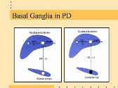

Functional Neurosurgery and Anesthetic Considerations
By: drdwayn Views: 1346
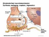
Neurotransmitter and Receptors
By: drdwayn Views: 1343
Chest Injuries
By: drdwayn Views: 2141
Anticoagulants
By: drdwayn Views: 2152
Routes of Bacterial Infection
By: drdwayn Views: 1251
Cadriovascular Disease Assessment
By: alaamadrid7 Views: 965
Blood Pressure Variability: The Good And The Bad
By: JenniferDwayne Views: 1697
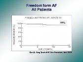
RF Ablation of Atrial Fibrillation in Valvular Heart Surgery Patients
By: yourdoctors Views: 352
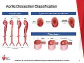
Guideline for the Diagnosis and Management of Aortic Disease
By: yourdoctors Views: 421
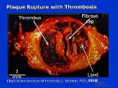
Pathogenesis of Acute Coronary Syndromes
By: JenniferDwayne Views: 1269

- About : I am Dr. Dwayne Faulk
- Occupation : Medical Professional
- Specialty : Other Health Professionals
- Country : United States of America
HEALTH A TO Z
- Eye Disease
- Heart Attack
- Medications
Home PowerPoint Templates Template Backgrounds Circulatory System PowerPoint Template
Circulatory System PowerPoint Template
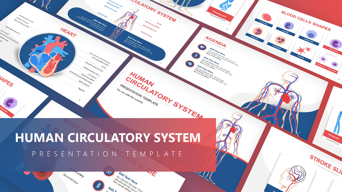
The Circulatory System PowerPoint Template is a health literacy presentation deck. It offers a collection of diagrams featuring functions of the cardiovascular system. These diagrams include the anatomy of blood vessels in the human body, their structure, arteries of heart, and cells. There are additional three slides of myocardial infarction, stroke, and varicose veins for heart conditions including structural problems and blood clots.
The circulatory system known as cardiovascular or vascular is an organ system of blood circulation in the body. It is made up of a network of blood vessels that transport blood to and from the heart. The blood vessels that carry blood away from the heart are called arteries and veins take the blood back to the heart. This system of transporting blood carries oxygen, nutrients, and hormones to cells.
The PowerPoint templates of Circulatory system diagrams are educational presentations. These templates can help communicate hearth health concepts to medical students or create presentations on specific topics such as a myocardial infarction PPT. The label diagrams of blood vessels and hearth display the components in a clear manner. The health professionals benefit from presentations of the cardiovascular system and related topics to discuss new medical developments. Users can copy these diagrams propose a convincing case for treatments or medical devices to venture capitalists and investors.
The Circulatory System PowerPoint Template is designed with vector images to illustrate the anatomy of the human heart. The vector-based graphics let users customize PowerPoint shapes by changing colors, sizes, and shape effects. Users benefit from editable graphics to recreate more high-quality diagrams by combining graphics from multiple slides. The slides of cell types are reusable in many medical presentations about healthy blood cells or diseases.
You must be logged in to download this file.
Favorite Add to Collection
Details (9 slides)

Supported Versions:
Subscribe today and get immediate access to download our PowerPoint templates.
Related PowerPoint Templates

Incident Report PowerPoint Template

Neuroscience PowerPoint Template
Cardiology PowerPoint Template

Human Growth & Development PowerPoint Template
The Circulatory System powerpoint
All Formats
Resource types, all resource types.
- Rating Count
- Price (Ascending)
- Price (Descending)
- Most Recent
The circulatory system powerpoint
The Circulatory System Activities Bundle: Reading Passages Worksheets PowerPoint

The Circulatory System Powerpoint

The Circulatory System Power Point Presentation
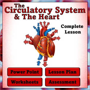
The Circulatory System and The Heart - Complete Lesson, PPT , Printables and Quiz

- Easel Assessment
Blood and Stuff -- The Circulatory System PowerPoint

The Circulatory System : Interactive Google Slides + PPT + Worksheet

- Google Apps™

BUNDLE- THE CIRCULATORY SYSTEM : Google Slides and creative revision activities

Introduction to the Circulatory System - Slides & Pear Deck Presentation

- Google Slides™
INTRODUCTION TO THE CIRCULATORY SYSTEM PPT AND ACTIVITIES

The Circulatory System - Middle School Science - Google Slides and Guided Notes

- Google Drive™ folder
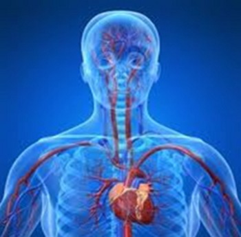
The Circulatory System - PowerPoint

Parts and Functions of The Circulatory System , Human Body Systems, Google Slides

- Internet Activities
THE CIRCULATORY SYSTEM : Google Slides and Activities
The Circulatory System PowerPoint

Parts Of The Circulatory System : Drag & Drop Worksheet: Google Slides . Powerpoint
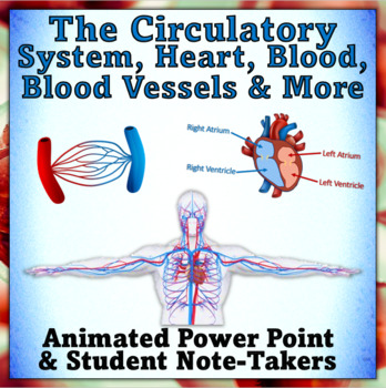
The Circulatory System - Power Point Presentation and Note-Taker
Introduction to the Circulatory System - Google Slides Presentation
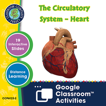
The Circulatory System – Heart - Google Slides Gr. 5-8

The Circulatory System – Blood - Google Slides Gr. 5-8
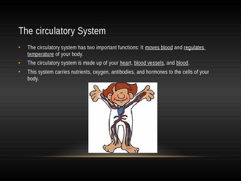
The Circulatory System the Complete Lesson Bundle ( Powerpoint and Assessments)

Biology 20: The Circulatory System Lesson Slides

Injuries, Defect, Disease, Disorder of the Circulatory System : GOOGLE SLIDES

The Circulatory System - Health Power Point

- We're hiring
- Help & FAQ
- Privacy policy
- Student privacy
- Terms of service
- Tell us what you think
The Circulatory System - PowerPoint
The Circulatory System is a fundamental topic covered in science education, and a PowerPoint presentation by Teacher of Primary offers an engaging resource to help students understand this vital bodily function. The circulatory system comprises the heart, blood vessels, and blood, each playing a crucial role in sustaining life. The heart, a muscle located centrally in the chest slightly towards the left and roughly the size of one's fist, is responsible for pumping blood throughout the body via blood vessels. This system's primary function is to circulate oxygen-rich blood and nutrients to various body parts while also removing waste products.
The heart operates as a dual-sided pump, with the left side receiving oxygenated blood from the lungs and sending it out to the body, while the right side collects deoxygenated blood from the body and pumps it back to the lungs for re-oxygenation. Structurally, the heart is divided into four chambers: the right and left atria at the top, which receive returning blood, and the right and left ventricles at the bottom, which are tasked with pumping blood out. Blood flow through the heart is meticulously controlled by the contraction of these chambers and the function of valves that prevent backflow, creating the familiar heartbeat sound. Blood vessels, including arteries, veins, and capillaries, facilitate the circulation of blood, and the pulse rate, typically between 70-100 beats per minute at rest, can be felt where arteries run close to the skin. The blood itself is a mixture of red blood cells, platelets, and white blood cells suspended in plasma, each with specific functions such as oxygen transport, clotting, and immune defense. The presentation also includes a quiz to test students' understanding of the circulatory system and its components.

Researched by Consultants from Top-Tier Management Companies

Powerpoint Templates
Icon Bundle
Kpi Dashboard
Professional
Business Plans
Swot Analysis
Gantt Chart
Business Proposal
Marketing Plan
Project Management
Business Case
Business Model
Cyber Security
Business PPT
Digital Marketing
Digital Transformation
Human Resources
Product Management
Artificial Intelligence
Company Profile
Acknowledgement PPT
PPT Presentation
Reports Brochures
One Page Pitch
Interview PPT
All Categories
Matters of the Cardiovascular System: Top 8 Templates to Know Its Mystery [Free PDF Attached]
![powerpoint presentation about the circulatory system Matters of the Cardiovascular System: Top 8 Templates to Know Its Mystery [Free PDF Attached]](https://www.slideteam.net/wp/wp-content/uploads/2022/08/1013x441no-button-8-1013x441.jpg)
Lakshya Khurana
From time immemorial, the human heart has drawn the fascination of love-struck poets. Considered the seat of pure human emotions like love, empathy, and kindness, it has been used to stir our emotions. Movies are one industry where matters of the heart feature the most often.
In this blog, we offer you the full love and kindness of the human heart, of course. Our key aim, however, is a bit more practical and down-to-earth.
At SlideTeam, we will talk about how the heart works (aka the cardiovascular or circulatory systems).
This blog post offers you a collection of eight PowerPoint sets from which you can choose. This collection comprises complete decks for the cardiovascular system with the heart and the blood vessels in focus.
Whether you are a high school student, a med student, or a doctor, it has no bearing on whether this collection is right for you because the templates are:
- CONTENT-READY: You do not need to do much research as we have curated that for you. Should you be running late, your presentation is already prepared.
- EDITABLE: Any information that catches your attention can be added to or deleted from these slides.
Each deck comes with several slides that present the diagram with and without the labels and highlight individual organ parts, making for a comprehensive presentation! Let us start our tour!
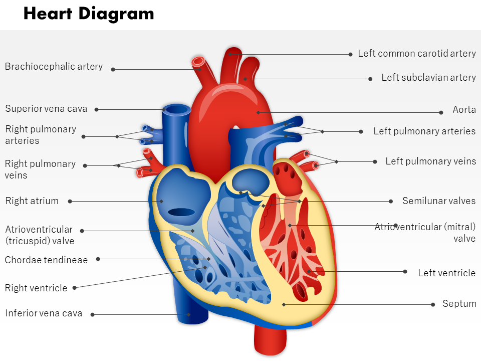
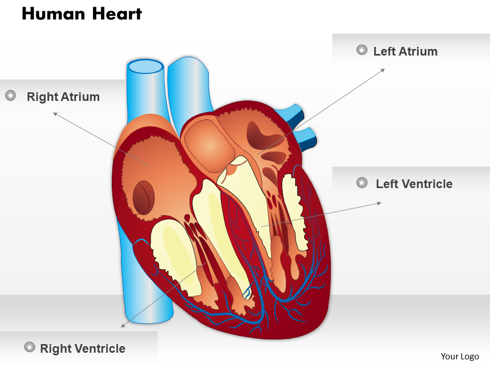
Template 1: Human Heart Anatomy PowerPoint Slides
This PPT layout presents a diagram of the human heart. Showcase color-coordinated figures of the heart and its components. Click on the link below to download this PowerPoint Preset.
Download this template
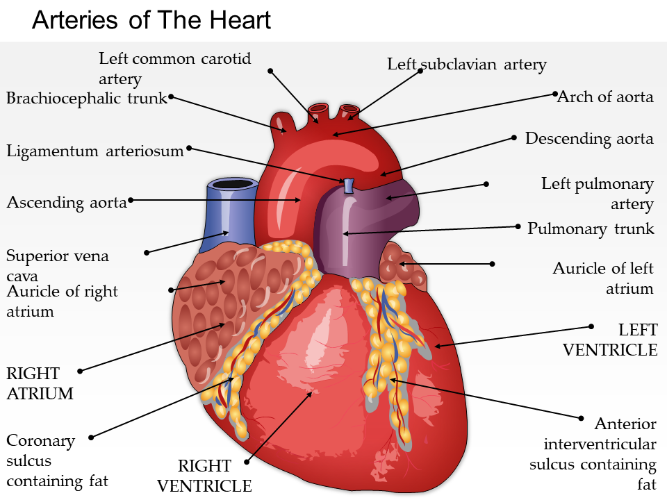
Template 2: Blood Vessels of the Heart
You’ve seen the blood vessels that run in your body, on your hands and feet. The blood vessels that run to and from the heart are quite special and large. Their roles, too, are different from what most of us presume. Do you know your stuff, as far as the heart is concerned? Employ this PowerPoint bundle to show your audience how.
Grab this template
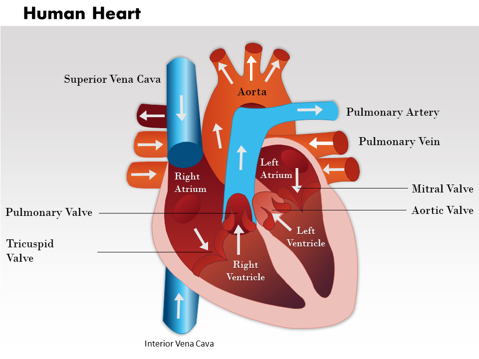
Template 3: Human Heart Cross-Section
This PPT set details the parts inside the heart, including the chambers, valves (which make the sound of the heartbeat), and blood vessels. Get this PowerPoint Preset to track your blood’s route with every heartbeat. No other company can give this feeling of being a Sherlock Homes or Hercules Poirot.
Template 4: Human Heart Musculature
Running for a few hours would turn our legs to jello. People often forget the heart is a muscle that does its work all day, every day, without stopping (over 100,000 beats a day). Present the heart’s musculature, muscle fibers, and inner components using this PowerPoint Deck.
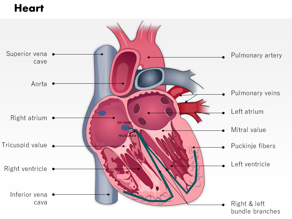

Template 5: Electrical System of the Heart
Besides love, it is electricity that keeps the heart going. That is why older people or people with heart problems require pacemakers and why doctors use electrical pads to resuscitate patients (in real life and in medical dramas). Use this PPT layout to showcase the wiring system that keeps the heart’s muscles working non-stop.
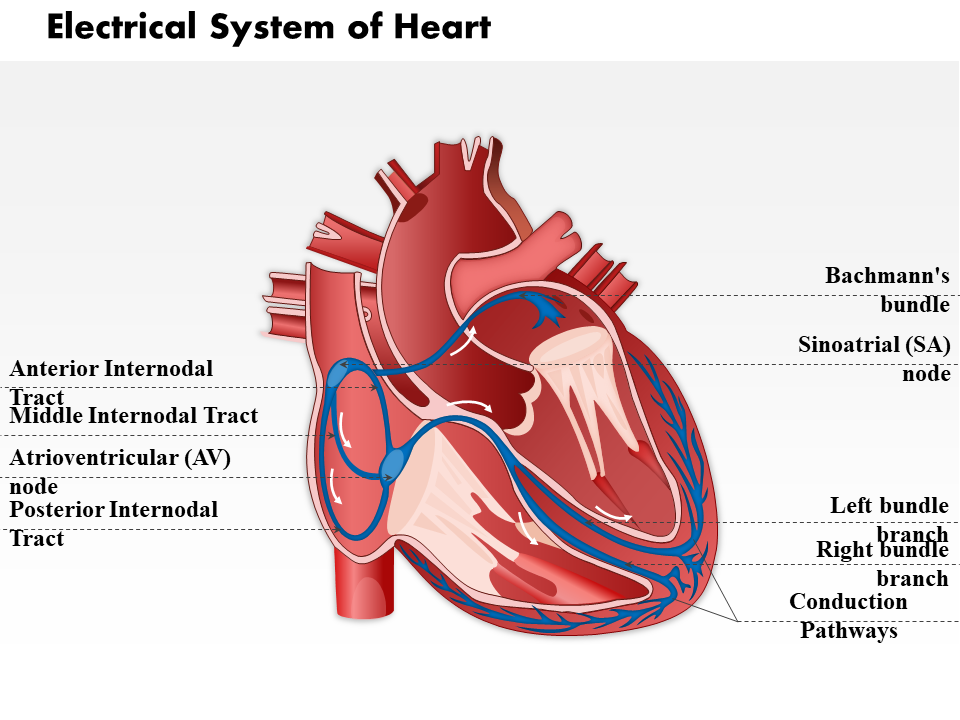
Template 6: Artery and Vein Diagrams
If you are lucky enough to see a blood vessel in a person, they actually look emaciated and eaten from inside. On the actual inside, however, they possess a complicated structure that ensures your oxygen and carbon dioxide go where they ought to. Detail this structure using this PowerPoint Theme. You may also scroll down for more information on blood vessels.
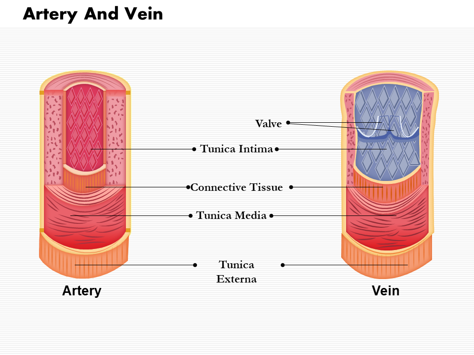
Template 7: The Heart’s Coronal Section
A coronal section is one that divides the body (in this case, the heart) into front and back, posterior and anterior. Use this PPT Set to present, in even greater detail, what lies within the human heart.
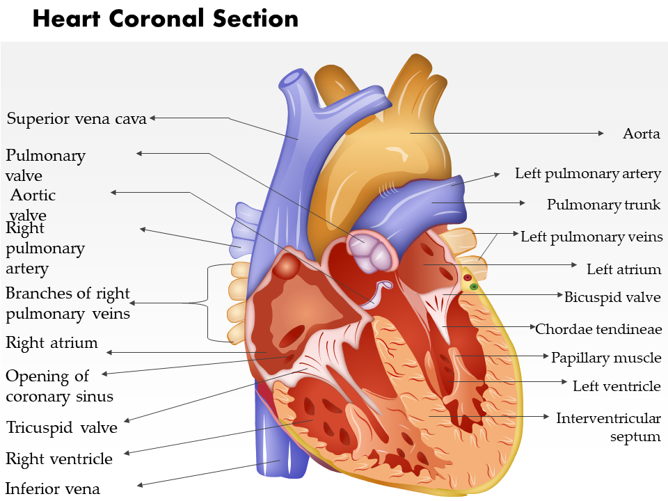
Template 8: The Human Heart’s Floor Plan
Your bedroom can’t be your living room or your kitchen your workspace. The heart has its own rooms, four, in fact, each separated by doors (valves) and with its specific purpose. Demonstrate which kind of blood resides in which chamber and why, with this PowerPoint Theme.
The Cardiovascular System: A Refresher
The following text is written to provide you with a refresher on the cardiovascular system, its function, and how it works. We'll cover everything from the heart and its chambers to the blood vessels and how they transport blood throughout the body. It has been written in a way to be understood by people of all educational background . By the end, it should help you brush up on your knowledge of the circulatory system and get you ready for your presentation.
The cardiovascular system comprises the heart, blood vessels, and blood. The heart is a muscular organ that pumps blood through the blood vessels to the rest of the body. The blood vessels are a network of tubes; the blood itself is a fluid that carries oxygen and nutrients to the body's cells and removes waste products from the cells.
The cardiovascular system has three main functions: transport oxygen and nutrients to the body's cells, remove waste products from the body's cells and maintain blood pressure. The circulatory system works with the respiratory system to provide oxygen to the cells and remove carbon dioxide from the body. It also works with the digestive system to absorb nutrients from food and eliminate waste products from the body.
The heart is a muscular organ that pumps blood (about 1.5 gallons per minute!) through the blood vessels to the rest of the body. The heart has four chambers: the right atrium, the left atrium, the right ventricle, and the left ventricle. The right atrium receives blood from the body and pumps it into the right ventricle. The right ventricle pumps blood into the lungs to pick up oxygen. The left atrium receives blood from the lungs and pumps it into the left ventricle. The left ventricle pumps blood out to the body.
The Blood Vessels
The blood vessels are a network of tubes that carry blood to and from the heart. There are three types of blood vessels: arteries, veins, and capillaries. Arteries carry blood away from the heart and have thick walls to withstand the high pressure of the blood. Veins carry blood back to the heart and have thin walls. Capillaries are tiny blood vessels that connect arteries and veins.
The blood comprises RBCs, white blood cells, platelets, and plasma. Red blood cells carry oxygen to the cells of the body. White blood cells fight infection. Platelets help the blood clot. Plasma is the liquid part of the blood that carries the red blood cells, white blood cells, and platelets.
The cardiovascular system is an important part of the body's systems and works to keep the body healthy.
1. What is the cardiovascular system?
The cardiovascular system is a network of organs and tissues that work together to move blood throughout the body. The heart pumps blood through the arteries, veins, and capillaries, which carry oxygen and nutrients to the cells and remove waste products.
2. What does the cardiovascular system do?
The cardiovascular system helps to regulate blood pressure, heart rate, and blood flow. It also helps to protect the body from infection and disease .
3. What are the main parts of the cardiovascular system?
The cardiovascular system comprises the heart, blood vessels, and blood.
4. What function do capillaries serve in the cardiovascular system?
Capillaries are the smallest of a body's blood vessels. The walls of capillaries are only one cell thick, which allows them to easily exchange oxygen and carbon dioxide between the blood and the tissues.
Download the free Cardiovascular System PDF .
Related posts:
- Questions Du Système Cardiovasculaire : Les 8 Meilleurs Modèles Pour Connaître Son Mystère
- Asuntos Del Sistema Cardiovascular: Top 8 Plantillas Para Conocer Su Misterio
- Angelegenheiten Des Herz-kreislauf-systems: Top 8 Vorlagen, Um Sein Geheimnis Zu Kennen
- Assuntos Do Sistema Cardiovascular: Os 8 Principais Modelos Para Conhecer Seu Mistério
Liked this blog? Please recommend us

[Updated 2023] The 5 Leadership Styles Along (With PPT Templates Included)

Top 15 Healthcare Brochure Templates to Advertize Your Medical Services
This form is protected by reCAPTCHA - the Google Privacy Policy and Terms of Service apply.

Digital revolution powerpoint presentation slides

Sales funnel results presentation layouts
3d men joinning circular jigsaw puzzles ppt graphics icons

Business Strategic Planning Template For Organizations Powerpoint Presentation Slides
Future plan powerpoint template slide

Project Management Team Powerpoint Presentation Slides

Brand marketing powerpoint presentation slides

Launching a new service powerpoint presentation with slides go to market

Agenda powerpoint slide show
Four key metrics donut chart with percentage

Engineering and technology ppt inspiration example introduction continuous process improvement
Meet our team representing in circular format

Got any suggestions?
We want to hear from you! Send us a message and help improve Slidesgo
Top searches
Trending searches
infertility
30 templates

linguistics
89 templates

15 templates

28 templates

public health
35 templates

holy spirit
38 templates
Cardiovascular Disease
Cardiovascular disease presentation, free google slides theme, powerpoint template, and canva presentation template.
It’s time to talk about cardiovascular diseases. Provide some data about them using this creative template full of illustrations of hearts, body diagrams, tables. We have also added useful sections to share data about the diagnosis, recommendations, pathology, treatment and processes.
Features of this template
- An abstract design with different hues of light pink, resembling blood
- 100% editable and easy to modify
- 25 different slides to impress your audience
- Contains easy-to-edit graphics such as tables, charts, diagrams and maps
- Includes 500+ icons and Flaticon’s extension for customizing your slides
- Designed to be used in Google Slides, Canva, and Microsoft PowerPoint
- 16:9 widescreen format suitable for all types of screens
- Includes information about fonts, colors, and credits of the free resources used
How can I use the template?
Am I free to use the templates?
How to attribute?
Attribution required If you are a free user, you must attribute Slidesgo by keeping the slide where the credits appear. How to attribute?
Related posts on our blog.

How to Add, Duplicate, Move, Delete or Hide Slides in Google Slides

How to Change Layouts in PowerPoint

How to Change the Slide Size in Google Slides
Related presentations.

Premium template
Unlock this template and gain unlimited access

HISTOLOGY OF THE CIRCULATORY SYSTEM
Jan 14, 2012
780 likes | 2.44k Views
HISTOLOGY OF THE CIRCULATORY SYSTEM. The cardiovascular system is subdivided into two functional parts Blood vascular system a. The blood vascular system distributes nutrients, gases, hormones to all parts of the body; collects wastes produced during cellular metabolism.
Share Presentation
- many blood vessels
- intrinsic contraction rate
- very similar
- lymph vascular
- squamous epithelium

Presentation Transcript
HISTOLOGY OF THE CIRCULATORY SYSTEM The cardiovascular system is subdivided into two functional parts Blood vascular system a. The blood vascular system distributes nutrients, gases, hormones to all parts of the body; collects wastes produced during cellular metabolism. b. The blood vascular system consists of a continuum of blood vessels (arteries, arterioles, capillaries, venules, veins) and a muscular pump (heart). c. Blood is the fluid found within the blood vascular system. 2. Lymph vascular system a. The lymph vascular system collects tissue fluid from tissues and returns it to the blood vascular system. b. The lymph vascular system consists of blind-ended capillaries (lymphatic capillaries) connected to venous vessels (lymphatic vessels) and various lymphoid organs (e.g., lymph nodes). c. The fluid found within the lymph vascular system is lymph. Composition of lymph in smaller lymphatic vessels is very similar to tissue fluid.
1. The heart wall can be viewed as a three-layered structure. • a. Inner layer = endocardium • b. Middle Layer = myocardium • c. Outer layer = epicardium (also called the pericardium) • 2. Except for the smallest vessels, blood and lymphatic vessel walls can also be viewed as • three-layered structures. • a. Inner layer = tunica intima • b. Middle layer = tunica media • c. Outer layer = tunica adventita
Structure of the heart wall • 1. The endocardium is the inner layer of the heart wall and consists of the endothelial • lining and the underlying connective tissue layers. • a. The lumen of the heart is lined by an endothelium consisting of a typical simple • squamous epithelium with well-developed zonulae occludens and basal lamina. • b. A connective tissue region consisting of three layers separates the endothelium from • the myocardium in humans consist of:- • (1) A thin layer of loose FECT (containing mainly fine collagen fibers) referred to as subendothelial layerwhih is next to the endothelium. • (2) A thicker layer of moderately dense FECT (with many elastic fibers) and some smooth muscle forms the center of the connective tissue region. • (3) A thin layer of loose FECT (often referred to as the subendocardial layer) • containing many blood vessels joins the endocardium to the myocardium • Purkinje fibers run in this layer in the interventricular septum.
2. The myocardium • is the middle layer of the heart wall and contains the cardiac muscle throughout most of the heart. • a. Cardiac muscle cells in the myocardium are arranged in strands whose ends attach to the dense connective tissue which surrounds the valves. • b. Loose FECT holds bundles of cardiac muscle cells/fibers together and contains numerous blood vessels. • c. Dense FECT (heavily collagenous) replaces the cardiac muscle in region around each of the major heart valves This connective tissue frame around each valve is called the cardiac skeleton
Cardiac Muscle Intercalated Disc
The epicardium • is the outer layer of the heart and consists of a connective tissue region covered by a mesothelium on its outer surface. • a. The connective tissue region consists of three layers in humans. • (1) The inner two regions are referred to collectively as the subepicardial layer and contain large blood vessels (coronary vessels), nerves, and varying amounts of adipose tissue. • (a) A thin layer of loose FECT lies next to the myocardium. • (b) A thicker layer of slightly denser FECT lies outside the loose FECT layer. • (2) A thin layer of loose FECT with many elastic fibers connects the connective tissue layers of the epicardium to the mesothelial covering.
DFIACT Adipose Tissue Coronary vessels and cardiac nerves Mesothelium Epicardium
b. A mesothelium (simple squamous epithelium) covers the outer surface of the heart (except where the arteries leave and the great veins enter the heart). This covering epithelium closely resembles the mesothelial covering of the other thoracic and abdominal organs. • B. The thickness of the heart wall and the thickness of the layers within the heart wall varies with location. • 1. The myocardium is thickest in the ventricular region, especially the left ventricle, and contains more cardiac muscle in the ventricles than in the atrium. The myocardium around the valves contains only dense collagenous CT which forms the cardiac skeleton. • 2. The endocardium and epicardium are thinner in the ventricles than in the atria • In the atria, the cardiac muscle cells contain small granules (called atrial specific granules) in the perinuclear sarcoplasm which can be observed with TEM. These granules are the source of atrial natriuretic peptide (ANP), a hormone which influences blood pressure by affecting kidney function
Special features of the heart • 1. Valves are out growths from the endocardium which prevent backflow • of blood. Valves contain three components. • . • 2. The cardiac skeleton supports each of the heart valves. Cardiac • muscle in the myocardium is replaced by dense regular FECT (heavily collagenous) • 3. Cardiac muscle fibers in the atria and ventricles are highly organized. • a. Cardiac muscle cells are attached end-to-end in branching strands. • b. The ends of most strands of cardiac muscle fibers are attached to the cardiac skeleton
"Pacemakers" • in the heart are modified cardiac muscle cells. • a. Cardiac muscle cells in the myocardium of the sinoatrial (SA) node are modified to serve as the pacemaker region. The plasma membrane of the cells has a high leakage rate, giving them the fastest intrinsic contraction rate among the populations • b. Cardiac muscle cells in the atrioventricular (AV) node have a similar histological appearance, but have a lower intrinsic rate of contraction, so these cells do not normally act as a pacemaker region. These cells receive the wave of excitation from the cardiac muscle of the atria and pass the excitation on to the bundle of His.
The impulse-conducting system • which connects the atria with the ventricles serves • several functions. • a. The impulse conducting system is made up of a series of Purkinje fibers which are specialized cardiac muscle cells. • (1) Purkinje fibers are organized into a branched bundle (Bundle of His) which • extends from the atrio-ventricular (AV) node, through the interventricular • septum down to the apex of the ventricles. • (2) Purkinje fibers are attached (by intercalated disks) to cardiac muscle cells in the • myocardium at the apex of the ventricles and along outer walls of both • ventricles • b. The impulse conducting system improves heart function in two ways
Conduction System = AV Bundle of His + Purkinje Fibers Purkinje Fibers Muscle
Microanatomy of Blood Vessels • Most larger blood vessel walls contain three major layers with sublayering. • 1. The tunica intima is the luminal layer. • a. The lumen is lined by an endothelium of simple squamous epithelium. • b. A subendothelial layer of loose FECT is present in most medium to large vessels • and may contain scattered smooth muscle in larger vessels. • 2. An internal elastic lamina (elastica interna) marks the boundary between the tunica • intima and the tunica media. • 3. The tunica media contains layers of either elastic laminae/lamellae (fenestrated sheets) or FECT alternating with layers of smooth muscle. • 4. If present, the external elastic lamina (elastica externa) marks the boundary between • the tunica media and the tunica adventita. • 5. The tunica adventita contains loose to moderately dense FECT, +/- scattered smooth • muscle cells. Small and medium arteries and veins are present in the tunica adventitia of large arteries and veins
Large arteries (also called elastic arteries or conducting arteries) • include the aorta and its largest main branches. • (a. Tunica intima - thin (relative to other layers in this type of vessel) • (1) Endothelium • (2) Subendothelial layer contains some smooth muscle, elastic fibers, collagen fibers • b. Internal elastic lamina - not as distinct as in other arteries • c. Tunica media - thick • (1) 40 - 60 distinct, concentrically arranged elastic laminae • (2) Between elastic laminae - fibroblasts, elastic fibers, collagen fibers, spiral (to circular) smooth muscle • d. Tunica adventita - thin; consists mainly of collagen fibers, blood vessels, nerves; some elastic fibers, fibroblasts, macrophages may also be present • 2. Function = to conduct blood from the heart to smaller arteries and to even out blood pressure and flow. The presence of elastic laminae gives these vessels elastic properties. They expand as the heart contracts (to modulate blood pressure and store energy) and recoil during ventricular relaxation (to maintain more even pressure in large arteries).
Medium to small arteries (also called muscular arteries) • Tunica intima - thin • (1) Endothelium • (2) Thin subendothelial layer consisting of scattered fine collagen and elastic fibers and a few fibroblasts • b. Internal elastic lamina - very distinct, usually folded • c. Tunica media - thick • (1) Circular smooth muscle, 5 - 40 layers • (2) Small amount of CT with collagen fibers and elastic fibers (longitudinal orientation) between muscle • (3) Thickness decreases as diameter of vessel decreases • d. External elastic lamina (May be indistinct in smaller muscular arteries) • e. Tunica adventita - thick; loose FECT • 2. Function - to distribute blood to smaller arterial vessels. The muscular wall resists damage due to relatively high blood pressure in these vessels
Arterioles • 1. Structure • a. Tunica intima - very thin consisting only of endothelium • b. Internal elastic lamina - usually present except in smaller arterioles • c. Tunica media - 1 to 5 layers of smooth muscle, some elastic fibers • d. Tunica adventita - thin, consisting of longitudinally arranged collagen and elastic • fibers • 2. Function - to redistribute blood flow to capillaries and to alter blood pressure by altering peripheral resistance to blood flow. Arterioles can change diameter very drastically therefore affecting blood pressure and flow patterns. Arterioles are referred to as peripheral resistance vessels.
Capillaries • 1. Structure - consist only of endothelium, but may be partially surrounded by pericytes. • Three types of capillaries may be distinguished • . • a. Continuous (type I) capillaries have relatively thick cytoplasm and • the capillary wall is continuous. Lateral cell surfaces of cells are characterized by • zonula occludens (tight junctions), so materials move across cells via pinocytosis or • diffusion. These capillaries occur in most organs. • b. Fenestrated (type II) capillaries (Figure 13.18) have extremely thin cytoplasm and • the capillary wall is perforated at intervals by pores or fenestrations. Lateral cell • surfaces are characterized by zonula occludens (tight junctions). Materials • apparently cross the cells through the fenestrations. These capillaries are found in • the kidney and in endocrine glands. • c. Sinusoidal capillaries are larger in diameter than the other types and have wide • spaces between the lateral edges of the adjacent endothelial cells, so materials • (and some cells) can move freely in and out of the capillary. Sinusoidal capillaries • are found in the spleen, liver, and bone marrow. • 2. Functions • a. Capillaries are the site of normal exchange of materials between blood and tissue • fluid. • b. Capillaries may be a site of exit of WBCs from blood into tissue under some conditions, although this is probably more frequent in venules.
Venules • Size varies from 10 microns (post-capillary venules) to 1 mm (muscular venules) • 2. Post-capillary venules • a. Structure - larger diameter than capillaries; consist of endothelium surrounded by pericytes • b. Functions • (1) Collect blood from capillaries • (2) Respond to vasoactive agents (e.g., histamine, serotonin) by altering permeability • (3) Also a site of exchange of materials between tissue fluid and blood • (4) Site of exit of WBCs from blood into tissue
Larger muscular venules • a. Structure • (1) Tunica intima - thin; endothelium surrounded by outer sheath of collagen fibers • (2) Tunica media - thin; 1 - 3 layers of smooth muscle (circular) with collagen and elastic fibers between muscles • (3) Tunica adventita - thick; loose FECT containing longitudinal collagen fibers and scattered elastic fibers and fibroblasts • b. Function - to collect blood from post-capillary venules
Small to medium veins • 1. Structure • a. Tunica intima - thin • (1) Endothelium • (2) Thin subendothelial layer • (3) May be folded to form valves • b. Tunica media - thin; circular smooth muscle, collagen fibers, some elastic fibers • c. Tunica adventita - well developed; loose FECT with longitudinally arranged collagen and elastic fibers, bundles of longitudinal smooth muscle • 2. Function - to collect blood from smaller venous vessels
Large veins - vena cavae and larger branches • 1. Structure • a. Tunica intima - thicker • (1) Endothelium • (2) Thin subendothelial layer • b. Internal elastic lamina - usually distinguishable • c. Tunica media - thin, poorly developed; mostly FECT; little smooth muscle • d. Tunica adventita - very thick; moderately dense FECT with spirally arranged collagen fibers, elastic laminae, longitudinal smooth muscle • 2. Function - to collect blood from medium sized veins and return it to heart
Microanatomy of Lymphatic Vessels • A. Lymph capillaries • 1. Structure - blind-ended tubules; consist only of endothelium (which lacks cell junctions); similar to post capillary venules of blood vascular system • 2. Function - to collect excess tissue fluid • B. Small to medium lymphatic vessels (Plate 31) • 1. Structure • (similar to venous blood vessels of the next smaller size) • a. Smaller lymphatic vessels consist of endothelium surrounded by collagen and elastic fibers and a few smooth muscle cells
Medium-sized lymphatic vessels • b. • (1) Tunica intima - thin; endothelium surrounded by few collagen and elastic fibers; may be folded to form valves • (2) Tunica media - thin; helically arranged smooth muscle, elastic fibers • (3) Tunica adventita - thicker; collagen and elastic fibers, few smooth muscle cells • 2. Function - to collect lymph from lymph capillaries
Large lymphatic vessels • C. include the thoracic duct and right lymphatic duct. • 1. Structure • a. Tunica intima - thin • (1) Endothelium • (2) Subendothelial layer of collagen and elastic fibers, some longitudinal smooth muscle • b. Tunica media - thickest; longitudinal and circular smooth muscle bundles, loose FECT (similar to a medium blood vein) • c. Tunica adventita - not well developed; coarse collagen fibers, few longitudinal smooth muscle • 2. Function - to collect lymph from medium sized lymphatic vessels and return it to largeveins • D. Lymphatic vessels of any size may appear empty, may contain faint pink material (proteins),or may contain lymphocytes.
- More by User
Histology of the Digestive System
Histology of the Digestive System. Basic Histological Layers Mucosa a. Epithelium b. Lamina Propria c. Muscularis Mucosae Submucosa a. Submucosal plexus “Plexus of Meissner” Muscularis a. Myenteric plexus “Plexus of Auerbach Serosa . Histology of the Mucosa.
1.91k views • 27 slides

Histology of the Nervous System
Histology of the Nervous System. Divisions of the Nervous System Anatomically CNS: Brain and Spinal cord PNS : Nerves and Ganglia Histologically Nerve cells Glial cells. Neurons Are the unit structure and function
611 views • 12 slides
The Circulatory System
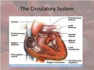
The Circulatory System. The Body’s Transport System. Like roads that link all parts of your town, your cardiovascular system links all the parts and systems of your body. In one year, your heart pumps enough blood to fill more than 30 swimming pools!
766 views • 45 slides

The Circulatory System. The Heart Facts. - The heart is a muscle. - The heart is a little larger than your clenched fist. - Your heart beats about 100,000 times in one day and about 35 million times in a year. During an average lifetime, the human heart will beat more than 2.5 billion times. .
733 views • 11 slides

The Circulatory System. The Heart. Heart: an organ made of cardiac muscle that pumps blood throughout the body Made of four parts called chambers The two upper chambers are called the left and right atriums The two lower chambers are called the left and right ventricles
323 views • 7 slides

The Circulatory System. May 18, 2012 pgs. 151-152. Know. What do you know about the circulatory System? . “I don’t know anything.” is not an acceptable answer. Use complete sentences. Evidence: Drawings pg 539 and 541 textbook– draw, label and color. Circulatory System. Functions.
280 views • 8 slides

The Circulatory System. By Yasheeka Patel, Amelia Norris, Lisa Ostman, Joyce Liu, Monica Macinante , Pabodha Gunaratne. FUNCTION: The Circulatory System is an organ system that allows blood to circulate and transport nutrients, oxygen, carbon dioxide, hormones, blood cells, and more.
189 views • 10 slides

The Circulatory System. Function: Transport system: Carries oxygen from the lungs to tissue cells throughout the body so they can do cell respiration Carries carbon dioxide, a waste product of cell respiration, from tissue cells to the lungs where it can be expelled
285 views • 16 slides

The Circulatory System. The Circulatory System. Heart Lungs Arteries Veins. BrainPop Circulatory system Heart. The Circulatory System. Blood brings OXYGEN and NUTRIENTS to organs and muscles. Blood removes waste from organs and muscles.
548 views • 12 slides

THE CIRCULATORY SYSTEM
THE CIRCULATORY SYSTEM. Temple College ECA. The Circulatory System. Function Components. The Heart. Circulation of the Heart. Electrical Conduction System. SA node AV node Bundle of his Bundle branches Purkinje network. Normal Heart Rates. Adult 60-80 beats/min
301 views • 9 slides

The Circulatory System. Parts and Function. The 3 main parts of The Circulatory system. The Heart The Blood Vessels Blood. THE HEART. Parts of The Heart. The Atria -Receiving Chambers The Ventricles -Pumping Chambers The Valves -Controls Flow The Septum
216 views • 11 slides

The Circulatory System. Round and round we go!. Your Blood – Fluid Transport. PLASMA!. Plasma is the fluid portion of the blood in which the blood cells move. It is straw-colored and makes up 55% of the total volume of blood!. Red Blood Cells: Oxygen Carriers. Round, disk-shaped cells
482 views • 27 slides

The Circulatory System. Natalie Janowiak, Anthony Storck, Emma Schwartz, Matt Gutt, Matt Gerber. Open Circulatory System. Blood directly bathes internal organs Hemolymph- body fluid, same as blood and interstitial fluid
334 views • 20 slides

The Circulatory System. The Heart. What is the point? Why do you have a heart? Made up of 4 chambers – 2 atria (right and left), and 2 ventricles (right and left).
238 views • 11 slides
Histology for Pathology Circulatory System
Histology for Pathology Circulatory System. Theresa Kristopaitis , MD Associate Professor Director of Mechanisms of Human Disease Kelli A. Hutchens, MD, FCAP Assistant Professor Assistant Director of Mechanisms of Human Disease Loyola Stritch School of Medicine. OBJECTIVES.
714 views • 20 slides

Histology of the digestive system
Histology of the digestive system. Gastrointestinal tract (GI tract). Mouth: Internal: nonkeratinized stratified squamous epithelium Middle: buccinator muscles, bone. 2. pharynx: skeletal muscle; nonkeratinized stratified squamous epithelium. 3. esophagus:
543 views • 12 slides
Histology of the Urinary System
Histology of the Urinary System. Lecture Objectives. Describe the normal microscopic appearance of the different parts of the kidney including cortex, medulla, juxtaglomerular apparatus and the distribution of the vasculature within the kidney.
463 views • 29 slides

239 views • 20 slides

HISTOLOGY OF THE URINARY SYSTEM
HISTOLOGY OF THE URINARY SYSTEM. Prof. Dr. Fauziah Othman Department of Human Anatomy Faculty of Medicine and Health Sciences Universiti Putra Malaysia. Contents. Kidney * Parts of nephron and structures Juxtaglomerular apparatus Functional aspects Blood circulation
601 views • 36 slides

The Circulatory System. The Heart, Blood Vessels, Blood Types. The Closed Circulatory System. Humans have a closed circulatory system , typical of all vertebrates, in which blood is confined to vessels and is distinct from the interstitial fluid.
386 views • 22 slides

The Circulatory System. An Overview of the Circulatory System. Check out how the heart works on the Web!! How the Heart Works. Where the Heart is located. Circulatory or Cardiovascular System.
413 views • 34 slides
Histology of the Endocrine System
Histology of the Endocrine System. Dr. Ahmed Abdulhussein AL-Huchami. Endocrine system is the system that regulates the tissue activities by a secretory product (hormone) Hormones are chemicals that are released ( from an endocrine cell ) in a small amount ,
742 views • 25 slides
- International
- Schools directory
- Resources Jobs Schools directory News Search

The Circulatory System - PowerPoint presentation and worksheet
Subject: Primary science
Age range: 7-11
Resource type: Other
Last updated
25 September 2023
- Share through email
- Share through twitter
- Share through linkedin
- Share through facebook
- Share through pinterest

Tes paid licence How can I reuse this?
Your rating is required to reflect your happiness.
It's good to leave some feedback.
Something went wrong, please try again later.
Very nice PowerPoint! There is, however, an error. On one slide it states that arteries carry oxygenated blood and veins carry deoxygenated blood. This is incorrect as pulmonary arteries carry deoxygenated blood to the lungs and pulmonary veins carry oxygenated blood to the heart. Arteries carry blood away from the heart, veins carry blood to the heart.
Empty reply does not make any sense for the end user
Report this resource to let us know if it violates our terms and conditions. Our customer service team will review your report and will be in touch.
Not quite what you were looking for? Search by keyword to find the right resource:

IMAGES
VIDEO
COMMENTS
Jun 17, 2013 •. 500 likes • 368,102 views. I. itutor. Education Health & Medicine Business. 1 of 32. Download Now. Download to read offline. The Circulatory System - Download as a PDF or view online for free.
The Circulatory System delivers food and oxygen to body cells and carries carbon dioxide and other waste products away from body cells. ... Presentation Project; Schedule: Tuesday: Discuss expectations. Wednesday: Work day with Chromebooks. Thursday: Work day with Chromebooks. Friday: Presentations. 20 of 21.
Blood travels from the heart to the body through blood vessels . There are 3 types of blood vessels : Download Presentation. white blood cell. circulatory system. lymphocytes fight. artery. atrium mitral valve tricuspid. white blood cells.
1 Cardiovascular (Circulatory) System 2 3 Systemic Circulation - delivers blood to all body cells and carries away waste Pulmonary Circulation - eliminates carbon dioxide and oxygenates blood (lung pathway) 4 Structure of the Heart Heart Size - about 14 cm x 9 cm (the size of a fist). Located in the mediastinum. between the 2 nd rib and ...
The Circulatory System. Interesting Facts. The heartbeat is strong enough to squirt blood 30 feet. The longer a boy's ring finger is, the less likely they are to have a heart attack (according to one study) The human heart beats ~35 million times per year. The heart pumps ~1,000,000 barrels of blood in a lifetime.
Slide 1-. The Circulatory System is the main cooling and transportation system for the human body has about 5 liters of blood continuously traveling through it Is composed of the heart, lungs, and blood vessels has three different parts: pulmonary circulation (lungs) coronary circulation (heart) systemic circulation, (the rest of the system's ...
Learn about the structure and function of the circulatory system in this PowerPoint presentation by Dr. Kim Bledsoe, a biology professor at Western Oregon University. You will find diagrams, animations, and explanations of the major components and processes of the circulatory system, such as the heart, blood vessels, blood, and lymphatic system.
The Circulatory System PowerPoint Template is a health literacy presentation deck. It offers a collection of diagrams featuring functions of the cardiovascular system. These diagrams include the anatomy of blood vessels in the human body, their structure, arteries of heart, and cells. There are additional three slides of myocardial infarction ...
pptx, 5.55 MB. pdf, 7.69 MB. This interactive PowerPoint covers the function of the circulatory system, advantages of double circulation, the structures of the heart and the structure and function of blood vessels. Activities and practice questions are included within the PowerPoint along with model answers. Recommended time: 2-4 lessons.
This is a 13 slide powerpoint which teaches about the circulatory system. It teaches the three main parts of the system, the three different kinds of blood vessels, and what is in blood. Subjects: Anatomy, General Science, Science. Grades: 2 nd - 5 th. Types: PowerPoint Presentations. $3.00.
muscle around the heart (myocardium) helps pump blood through circulatory system. -. as blood flows through the system, it travels through three vessels (arteries, capillaries, and veins) -. arteries carry blood from the heart to tissues. -. capillaries bring nutrients to tissues and absorb carbon dioxide and waste. -.
Human Circulatory System. Human Circulatory Organs And Blood Circulation. Blood Vessel. Blood vessel is the organ that functions carrying blood to come out or come into heart. Human has three blood vessels which are artery, capillary, and vein. ARTERY. Slideshow 3077586 by orde.
Year 6 science programme of study - Animals, including humans: identify and name the main parts of the human circulatory system, and describe the functions of the heart, blood vessels and blood In this science teaching resource, pupils will learn about the main parts of the circulatory system and how it moves blood around the body. 'The ...
Template 1: Human Heart Anatomy PowerPoint Slides This PPT layout presents a diagram of the human heart. Showcase color-coordinated figures of the heart and its components. Click on the link below to download this PowerPoint Preset. ... The circulatory system works with the respiratory system to provide oxygen to the cells and remove carbon ...
Free Google Slides theme, PowerPoint template, and Canva presentation template. It's time to talk about cardiovascular diseases. Provide some data about them using this creative template full of illustrations of hearts, body diagrams, tables. We have also added useful sections to share data about the diagnosis, recommendations, pathology ...
In the Circulatory System, the heart, lungs, and blood vessels have to work together to maintain homeostasis. The Heart: pumps oxygen rich blood, nutrients, hormones, and other things the body needs to maintain health, to organs and tissues. Parts of the Heart The heart is made up of 4 different blood-filled areas.
The cardiovascular system is subdivided into two functional parts Blood vascular system a. The blood vascular system distributes nutrients, gases, hormones to all parts of the body; collects wastes produced during cellular metabolism. Download Presentation. only dense. many blood vessels. intrinsic contraction rate. very similar. lymph vascular.
ppt, 6.9 MB. pdf, 103.2 KB. In this teaching resource pupils will be taught to identify and name the main parts of the circulatory system, and describe the functions of the heart, blood vessels and blood. It contains a 35 slide PowerPoint presentation and 2 worksheets. This science teaching resource is an ideal tool to use in a lesson covering ...