- Open access
- Published: 15 April 2024

Demuxafy : improvement in droplet assignment by integrating multiple single-cell demultiplexing and doublet detection methods
- Drew Neavin 1 ,
- Anne Senabouth 1 ,
- Himanshi Arora 1 , 2 ,
- Jimmy Tsz Hang Lee 3 ,
- Aida Ripoll-Cladellas 4 ,
- sc-eQTLGen Consortium ,
- Lude Franke 5 ,
- Shyam Prabhakar 6 , 7 , 8 ,
- Chun Jimmie Ye 9 , 10 , 11 , 12 ,
- Davis J. McCarthy 13 , 14 ,
- Marta Melé 4 ,
- Martin Hemberg 15 &
- Joseph E. Powell ORCID: orcid.org/0000-0002-5070-4124 1 , 16
Genome Biology volume 25 , Article number: 94 ( 2024 ) Cite this article
Metrics details
Recent innovations in single-cell RNA-sequencing (scRNA-seq) provide the technology to investigate biological questions at cellular resolution. Pooling cells from multiple individuals has become a common strategy, and droplets can subsequently be assigned to a specific individual by leveraging their inherent genetic differences. An implicit challenge with scRNA-seq is the occurrence of doublets—droplets containing two or more cells. We develop Demuxafy, a framework to enhance donor assignment and doublet removal through the consensus intersection of multiple demultiplexing and doublet detecting methods. Demuxafy significantly improves droplet assignment by separating singlets from doublets and classifying the correct individual.
Droplet-based single-cell RNA sequencing (scRNA-seq) technologies have provided the tools to profile tens of thousands of single-cell transcriptomes simultaneously [ 1 ]. With these technological advances, combining cells from multiple samples in a single capture is common, increasing the sample size while simultaneously reducing batch effects, cost, and time. In addition, following cell capture and sequencing, the droplets can be demultiplexed—each droplet accurately assigned to each individual in the pool [ 2 , 3 , 4 , 5 , 6 , 7 ].
Many scRNA-seq experiments now capture upwards of 20,000 droplets, resulting in ~16% (3,200) doublets [ 8 ]. Current demultiplexing methods can also identify doublets—droplets containing two or more cells—from different individuals (heterogenic doublets). These doublets can significantly alter scientific conclusions if they are not effectively removed. Therefore, it is essential to remove doublets from droplet-based single-cell captures.
However, demultiplexing methods cannot identify droplets containing multiple cells from the same individual (homogenic doublets) and, therefore, cannot identify all doublets in a single capture. If left in the dataset, those doublets could appear as transitional cells between two distinct cell types or a completely new cell type. Accordingly, additional methods have been developed to identify heterotypic doublets (droplets that contain two cells from different cell types) by comparing the transcriptional profile of each droplet to doublets simulated from the dataset [ 9 , 10 , 11 , 12 , 13 , 14 , 15 ]. It is important to recognise that demultiplexing methods achieve two functions—segregation of cells from different donors and separation of singlets from doublets—while doublet detecting methods solely classify singlets versus doublets.
Therefore, demultiplexing and transcription-based doublet detecting methods provide complementary information to improve doublet detection, providing a cleaner dataset and more robust scientific results. There are currently five genetic-based demultiplexing [ 2 , 3 , 4 , 5 , 6 , 7 , 16 ] and seven transcription-based doublet-detecting methods implemented in various languages [ 9 , 10 , 11 , 12 , 13 , 14 , 15 ]. Under different scenarios, each method is subject to varying performance and, in some instances, biases in their ability to accurately assign cells or detect doublets from certain conditions. The best combination of methods is currently unclear but will undoubtedly depend on the dataset and research question.
Therefore, we set out to identify the best combination of genetic-based demultiplexing and transcription-based doublet-detecting methods to remove doublets and partition singlets from different donors correctly. In addition, we have developed a software platform ( Demuxafy ) that performs these intersectional methods and provides additional commands to simplify the execution and interpretation of results for each method (Fig. 1 a).
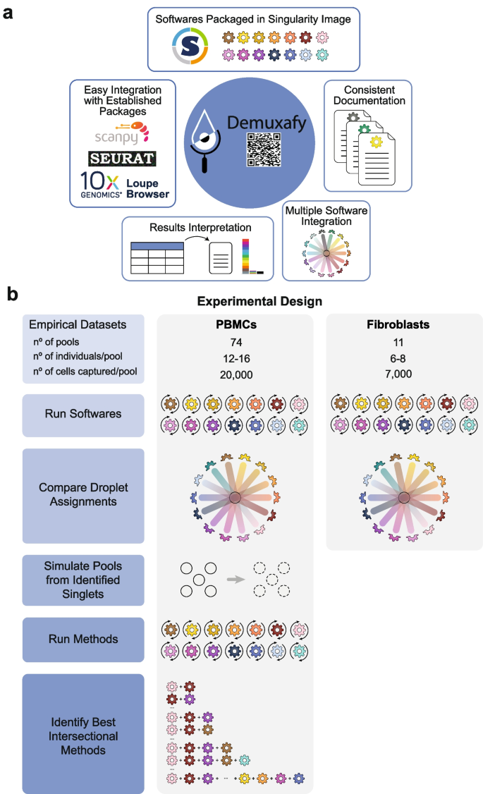
Study design and qualitative method classifications. a Demuxafy is a platform to perform demultiplexing and doublet detecting with consistent documentation. Demuxafy also provides wrapper scripts to quickly summarize the results from each method and assign clusters to each individual with reference genotypes when a reference-free demultiplexing method is used. Finally, Demuxafy provides a script to easily combine the results from multiple different methods into a single data frame and it provides a final assignment for each droplet based on the combination of multiple methods. In addition, Demuxafy provides summaries of the number of droplets classified as singlets or doublets by each method and a summary of the number of droplets assigned to each individual by each of the demultiplexing methods. b Two datasets are included in this analysis - a PBMC dataset and a fibroblast dataset. The PBMC dataset contains 74 pools that captured approximately 20,000 droplets each with 12-16 donor cells multiplexed per pool. The fibroblast dataset contains 11 pools of roughly 7,000 droplets per pool with sizes ranging from six to eight donors per pool. All pools were processed by all demultiplexing and doublet detecting methods and the droplet and donor classifications were compared between the methods and between the PBMCs and fibroblasts. Then the PBMC droplets that were classified as singlets by all methods were taken as ‘true singlets’ and used to generate new pools in silico. Those pools were then processed by each of the demultiplexing and doublet detecting methods and intersectional combinations of demultiplexing and doublet detecting methods were tested for different experimental designs
To compare the demultiplexing and doublet detecting methods, we utilised two large, multiplexed datasets—one that contained ~1.4 million peripheral blood mononuclear cells (PBMCs) from 1,034 donors [ 17 ] and one with ~94,000 fibroblasts from 81 donors [ 18 ]. We used the true singlets from the PBMC dataset to generate new in silico pools to assess the performance of each method and the multi-method intersectional combinations (Fig. 1 b).
Here, we compare 14 demultiplexing and doublet detecting methods with different methodological approaches, capabilities, and intersectional combinations. Seven of those are demultiplexing methods ( Demuxalot [ 6 ], Demuxlet [ 3 ], Dropulation [ 5 ], Freemuxlet [ 16 ], ScSplit [ 7 ], Souporcell [ 4 ], and Vireo [ 2 ]) which leverage the common genetic variation between individuals to identify cells that came from each individual and to identify heterogenic doublets. The seven remaining methods ( DoubletDecon [ 9 ], DoubletDetection [ 14 ], DoubletFinder [ 10 ], ScDblFinder [ 11 ], Scds [ 12 ], Scrublet [ 13 ], and Solo [ 15 ]) identify doublets based on their similarity to simulated doublets generated by adding the transcriptional profiles of two randomly selected droplets in the dataset. These methods assume that the proportion of real doublets in the dataset is low, so combining any two droplets will likely represent the combination of two singlets.
We identify critical differences in the performance of demultiplexing and doublet detecting methods to classify droplets correctly. In the case of the demultiplexing techniques, their performance depends on their ability to identify singlets from doublets and assign a singlet to the correct individual. For doublet detecting methods, the performance is based solely on their ability to differentiate a singlet from a doublet. We identify limitations in identifying specific doublet types and cell types by some methods. In addition, we compare the intersectional combinations of these methods for multiple experimental designs and demonstrate that intersectional approaches significantly outperform all individual techniques. Thus, the intersectional methods provide enhanced singlet classification and doublet removal—a critical but often under-valued step of droplet-based scRNA-seq processing. Our results demonstrate that intersectional combinations of demultiplexing and doublet detecting software provide significant advantages in droplet-based scRNA-seq preprocessing that can alter results and conclusions drawn from the data. Finally, to provide easy implementation of our intersectional approach, we provide Demuxafy ( https://demultiplexing-doublet-detecting-docs.readthedocs.io/en/latest/index.html ) a complete platform to perform demultiplexing and doublet detecting intersectional methods (Fig. 1 a).
Study design
To evaluate demultiplexing and doublet detecting methods, we developed an experimental design that applies the different techniques to empirical pools and pools generated in silico from the combination of true singlets—droplets identified as singlets by every method (Fig. 1 a). For the first phase of this study, we used two empirical multiplexed datasets—the peripheral blood mononuclear cell (PBMC) dataset containing ~1.4 million cells from 1034 donors and a fibroblast dataset of ~94,000 cells from 81 individuals (Additional file 1 : Table S1). We chose these two cell systems to assess the methods in heterogeneous (PBMC) and homogeneous (fibroblast) cell types.
Demultiplexing and doublet detecting methods perform similarly for heterogeneous and homogeneous cell types
We applied the demultiplexing methods ( Demuxalot , Demuxlet , Dropulation , Freemuxlet , ScSplit , Souporcell , and Vireo ) and doublet detecting methods ( DoubletDecon , DoubletDetection , DoubletFinder , ScDblFinder , Scds , Scrublet , and Solo ) to the two datasets and assessed the results from each method. We first compared the droplet assignments by identifying the number of singlets and doublets identified by a given method that were consistently annotated by all methods (Fig. 2 a–d). We also identified the percentage of droplets that were annotated consistently between pairs of methods (Additional file 2 : Fig S1). In the cases where two demultiplexing methods were compared to one another, both the droplet type (singlet or doublet) and the assignment of the droplet to an individual had to match to be considered in agreement. In all other comparisons (i.e. demultiplexing versus doublet detecting and doublet detecting versus doublet detecting), only the droplet type (singlet or doublet) was considered for agreement since doublet detecting methods cannot annotate donor assignment. We found that the two method types were more similar to other methods of the same type (i.e., demultiplexing versus demultiplexing and doublet detecting versus doublet detecting) than they were to methods from a different type (demultiplexing methods versus doublet detecting methods; Supplementary Fig 1). We found that the similarity of the demultiplexing and doublet detecting methods was consistent in the PBMC and fibroblast datasets (Pearson correlation R = 0.78, P -value < 2×10 −16 ; Fig S1a-c). In addition, demultiplexing methods were more similar than doublet detecting methods for both the PBMC and fibroblast datasets (Wilcoxon rank-sum test: P < 0.01; Fig. 2 a–b and Additional file 2 : Fig S1).
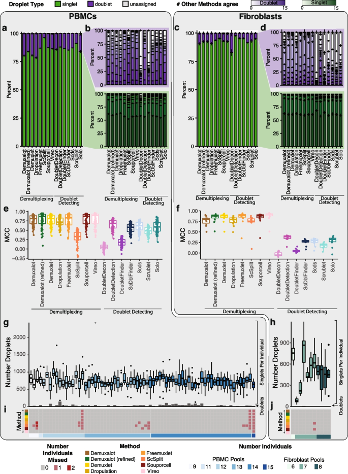
Demultiplexing and Doublet Detecting Method Performance Comparison. a The proportion of droplets classified as singlets and doublets by each method in the PBMCs. b The number of other methods that classified the singlets and doublets identified by each method in the PBMCs. c The proportion of droplets classified as singlets and doublets by each method in the fibroblasts. d The number of other methods that classified the singlets and doublets identified by each method in the fibroblasts. e - f The performance of each method when the majority classification of each droplet is considered the correct annotation in the PBMCs ( e ) and fibroblasts ( f ). g - h The number of droplets classified as singlets (box plots) and doublets (bar plots) by all methods in the PBMC ( g ) and fibroblast ( h ) pools. i - j The number of donors that were not identified by each method in each pool for PBMCs ( i ) and fibroblasts ( j ). PBMC: peripheral blood mononuclear cell. MCC: Matthew’s correlationcoefficient
The number of unique molecular identifiers (UMIs) and genes decreased in droplets that were classified as singlets by a larger number of methods while the mitochondrial percentage increased in both PBMCs and fibroblasts (Additional file 2 : Fig S2).
We next interrogated the performance of each method using the Matthew’s correlation coefficient (MCC) to calculate the consistency between Demuxify and true droplet classification. We identified consistent trends in the MCC scores for each method between the PBMCs (Fig. 2 e) and fibroblasts (Fig. 2 f). These data indicate that the methods behave similarly, relative to one another, for heterogeneous and homogeneous datasets.
Next, we sought to identify the droplets concordantly classified by all demultiplexing and doublet detecting methods in the PBMC and fibroblast datasets. On average, 732 singlets were identified for each individual by all the methods in the PBMC dataset. Likewise, 494 droplets were identified as singlets for each individual by all the methods in the fibroblast pools. However, the concordance of doublets identified by all methods was very low for both datasets (Fig. 2 e–f). Notably, the consistency of classifying a droplet as a doublet by all methods was relatively low (Fig. 2 b,d,g, and h). This suggests that doublet identification is not consistent between all the methods. Therefore, further investigation is required to identify the reasons for these inconsistencies between methods. It also suggests that combining multiple methods for doublet classification may be necessary for more complete doublet removal. Further, some methods could not identify all the individuals in each pool (Fig. 2 i–j). The non-concordance between different methods demonstrates the need to effectively test each method on a dataset where the droplet types are known.
Computational resources vary for demultiplexing and doublet detecting methods
We recorded each method’s computational resources for the PBMC pools, with ~20,000 cells captured per pool (Additional file 1 : Table S1). Of the demultiplexing methods, ScSplit took the most time (multiple days) and required the most steps, but Demuxalot , Demuxlet , and Freemuxlet used the most memory. Solo took the longest time (median 13 h) and most memory to run for the doublet detecting methods but is the only method built to be run directly from the command line, making it easy to implement (Additional file 2 : Fig S3).
Generate pools with known singlets and doublets
However, there is no gold standard to identify which droplets are singlets or doublets. Therefore, in the second phase of our experimental design (Fig. 1 b), we used the PBMC droplets classified as singlets by all methods to generate new pools in silico. We chose to use the PBMC dataset since our first analyses indicated that method performance is similar for homogeneous (fibroblast) and heterogeneous (PBMC) cell types (Fig. 2 and Additional file 2 : Fig S1) and because we had many more individuals available to generate in silico pools from the PBMC dataset (Additional file 1 : Table S1).
We generated 70 pools—10 each of pools that included 2, 4, 8, 16, 32, 64, or 128 individuals (Additional file 1 : Table S2). We assume a maximum 20% doublet rate as it is unlikely researchers would use a technology that has a higher doublet rate (Fig. 3 a).
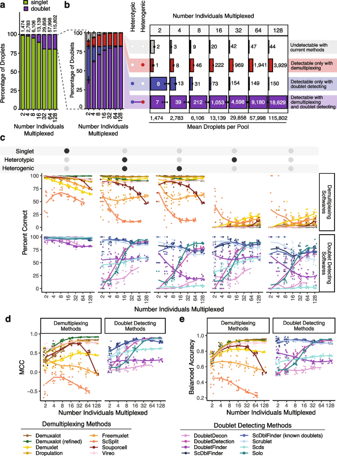
In silico Pool Doublet Annotation and Method Performance. a The percent of singlets and doublets in the in -silico pools - separated by the number of multiplexed individuals per pool. b The percentage and number of doublets that are heterogenic (detectable by demultiplexing methods), heterotypic (detectable by doublet detecting methods), both (detectable by either method category) and neither (not detectable with current methods) for each multiplexed pool size. c Percent of droplets that each of the demultiplexing and doublet detecting methods classified correctly for singlets and doublet subtypes for different multiplexed pool sizes. d Matthew’s Correlation Coefficient (MCC) for each of the methods for each of the multiplexed pool sizes. e Balanced accuracy for each of the methods for each of the multiplexed pool sizes
We used azimuth to classify the PBMC cell types for each droplet used to generate the in silico pools [ 19 ] (Additional file 2 : Fig S4). As these pools have been generated in silico using empirical singlets that have been well annotated, we next identified the proportion of doublets in each pool that were heterogenic, heterotypic, both, and neither. This approach demonstrates that a significant percentage of doublets are only detectable by doublet detecting methods (homogenic and heterotypic) for pools with 16 or fewer donors multiplexed (Fig. 3 b).
While the total number of doublets that would be missed if only using demultiplexing methods appears small for fewer multiplexed individuals (Fig. 3 b), it is important to recognise that this is partly a function of the ~732 singlet cells per individual used to generate these pools. Hence, the in silico pools with fewer individuals also have fewer cells. Therefore, to obtain numbers of doublets that are directly comparable to one another, we calculated the number of each doublet type that would be expected to be captured with 20,000 cells when 2, 4, 8, 16, or 32 individuals were multiplexed (Additional file 2 : Fig S5). These results demonstrate that many doublets would be falsely classified as singlets since they are homogenic when just using demultiplexing methods for a pool of 20,000 cells captured with a 16% doublet rate (Additional file 2 : Fig S5). However, as more individuals are multiplexed, the number of droplets that would not be detectable by demultiplexing methods (homogenic) decreases. This suggests that typical workflows that use only one demultiplexing method to remove doublets from pools that capture 20,000 droplets with 16 or fewer multiplexed individuals fail to adequately remove between 173 (16 multiplexed individuals) and 1,325 (2 multiplexed individuals) doublets that are homogenic and heterotypic which could be detected by doublet detecting methods (Additional file 2 : Fig S5). Therefore, a technique that uses both demultiplexing and doublet detecting methods in parallel will complement more complete doublet removal methods. Consequently, we next set out to identify the demultiplexing and doublet detecting methods that perform the best on their own and in concert with other methods.
Doublet and singlet droplet classification effectiveness varies for demultiplexing and doublet detecting methods
Demultiplexing methods fail to classify homogenic doublets.
We next investigated the percentage of the droplets that were correctly classified by each demultiplexing and doublet detecting method. In addition to the seven demultiplexing methods, we also included Demuxalot with the additional steps to refine the genotypes that can then be used for demultiplexing— Demuxalot (refined). Demultiplexing methods correctly classify a large portion of the singlets and heterogenic doublets (Fig. 3 c). This pattern is highly consistent across different cell types, with the notable exceptions being decreased correct classifications for erythrocytes and platelets when greater than 16 individuals are multiplexed (Additional file 2 : Fig S6).
However, Demuxalot consistently demonstrates the highest correct heterogenic doublet classification. Further, the percentage of the heterogenic doublets classified correctly by Souporcell decreases when large numbers of donors are multiplexed. ScSplit is not as effective as the other demultiplexing methods at classifying heterogenic doublets, partly due to the unique doublet classification method, which assumes that the doublets will generate a single cluster separate from the donors (Table 1 ). Importantly, the demultiplexing methods identify almost none of the homogenic doublets for any multiplexed pool size—demonstrating the need to include doublet detecting methods to supplement the demultiplexing method doublet detection.
Doublet detecting method classification performances vary greatly
In addition to assessing each of the methods with default settings, we also evaluated ScDblFinder with ‘known doublets’ provided. This method can take already known doublets and use them when detecting doublets. For these cases, we used the droplets that were classified as doublets by all the demultiplexing methods as ‘known doublets’.
Most of the methods classified a similarly high percentage of singlets correctly, with the exceptions of DoubletDecon and DoubletFinder for all pool sizes (Fig. 3 c). However, unlike the demultiplexing methods, there are explicit cell-type-specific biases for many of the doublet detecting methods (Additional file 2 : Fig S7). These differences are most notable for cell types with fewer cells (i.e. ASDC and cDC2) and proliferating cells (i.e. CD4 Proliferating, CD8 Proliferating, and NK Proliferating). Further, all of the softwares demonstrate high correct percentages for some cell types including CD4 Naïve and CD8 Naïve (Additional file 2 : Fig S7).
As expected, all doublet detecting methods identified heterotypic doublets more effectively than homotypic doublets (Fig. 3 c). However, ScDblFinder and Scrublet classified the most doublets correctly across all doublet types for pools containing 16 individuals or fewer. Solo was more effective at identifying doublets than Scds for pools containing more than 16 individuals. It is also important to note that it was not feasible to run DoubletDecon for the largest pools containing 128 multiplexed individuals and an average of 115,802 droplets (range: 113,594–119,126 droplets). ScDblFinder performed similarly when executed with and without known doublets (Pearson correlation P = 2.5 × 10 -40 ). This suggests that providing known doublets to ScDblFinder does not offer an added benefit.
Performances vary between demultiplexing and doublet detecting method and across the number of multiplexed individuals
We assessed the overall performance of each method with two metrics: the balanced accuracy and the MCC. We chose to use balanced accuracy since, with unbalanced group sizes, it is a better measure of performance than accuracy itself. Further, the MCC has been demonstrated as a more reliable statistical measure of performance since it considers all possible categories—true singlets (true positives), false singlets (false positives), true doublets (true negatives), and false doublets (false negatives). Therefore, a high score on the MCC scale indicates high performance in each metric. However, we provide additional performance metrics for each method (Additional file 1 : Table S3). For demultiplexing methods, both the droplet type (singlet or doublet) and the individual assignment were required to be considered a ‘true singlet’. In contrast, only the droplet type (singlet or doublet) was needed for doublet detection methods.
The MCC and balanced accuracy metrics are similar (Spearman’s ⍴ = 0.87; P < 2.2 × 10 -308 ). Further, the performance of Souporcell decreases for pools with more than 32 individuals multiplexed for both metrics (Student’s t -test for MCC: P < 1.1 × 10 -9 and balanced accuracy: P < 8.1 × 10 -11 ). Scds , ScDblFinder , and Scrublet are among the top-performing doublet detecting methods Fig. 3 d–e).
Overall, between 0.4 and 78.8% of droplets were incorrectly classified by the demultiplexing or doublet detecting methods depending on the technique and the multiplexed pool size (Additional file 2 : Fig S8). Demuxalot (refined) and DoubletDetection demonstrated the lowest percentage of incorrect droplets with about 1% wrong in the smaller pools (two multiplexed individuals) and about 3% incorrect in pools with at least 16 multiplexed individuals. Since some transitional states and cell types are present in low percentages in total cell populations (i.e. ASDCs at 0.02%), incorrect classification of droplets could alter scientific interpretations of the data, and it is, therefore, ideal for decreasing the number of erroneous assignments as much as possible.
False singlets and doublets demonstrate different metrics than correctly classified droplets
We next asked whether specific cell metrics might contribute to false singlet and doublet classifications for different methods. Therefore, we compared the number of genes, number of UMIs, mitochondrial percentage and ribosomal percentage of the false singlets and doublets to equal numbers of correctly classified cells for each demultiplexing and doublet detecting method.
The number of UMIs (Additional file 2 : Fig S9 and Additional file 1 : Table S4) and genes (Additional file 2 : Fig S10 and Additional file 1 : Table S5) demonstrated very similar distributions for all comparisons and all methods (Spearman ⍴ = 0.99, P < 2.2 × 10 -308 ). The number of UMIs and genes were consistently higher in false singlets and lower in false doublets for most demultiplexing methods except some smaller pool sizes (Additional file 2 : Fig S9a and Additional file 2 : Fig S10a; Additional file 1 : Table S4 and Additional file 1 : Table S5). The number of UMIs and genes was consistently higher in droplets falsely classified as singlets by the doublet detecting methods than the correctly identified droplets (Additional file 2 : Fig S9b and Additional file 2 : Fig S10b; Additional file 1 : Table S4 and Additional file 1 : Table S5). However, there was less consistency in the number of UMIs and genes detected in false singlets than correctly classified droplets between the different doublet detecting methods (Additional file 2 : Fig S9b and Additional file 2 : Fig S10b; Additional file 1 : Table S4 and Additional file 1 : Table S5).
The ribosomal percentage of the droplets falsely classified as singlets or doublets is similar to the correctly classified droplets for most methods—although they are statistically different for larger pool sizes (Additional file 2 : Fig S11a and Additional file 1 : Table S6). However, the false doublets classified by some demultiplexing methods ( Demuxalot , Demuxalot (refined), Demuxlet , ScSplit , Souporcell , and Vireo ) demonstrated higher ribosomal percentages. Some doublet detecting methods ( ScDblFinder , ScDblFinder with known doublets and Solo) demonstrated higher ribosomal percentages for the false doublets while other demonstrated lower ribosomal percentages ( DoubletDecon , DoubletDetection , and DoubletFinder ; Additional file 2 : Fig S11b and Additional file 1 : Table S6).
Like the ribosomal percentage, the mitochondrial percentage in false singlets is also relatively similar to correctly classified droplets for both demultiplexing (Additional file 2 : Fig S12a and Additional file 1 : Table S7) and doublet detecting methods (Additional file 2 : Fig S12b). The mitochondrial percentage for false doublets is statistically lower than the correctly classified droplets for a few larger pools for Freemuxlet , ScSplit , and Souporcell . The doublet detecting method Solo also demonstrates a small but significant decrease in mitochondrial percentage in the false doublets compared to the correctly annotated droplets. However, other doublet detecting methods including DoubletFinder and the larger pools of most other methods demonstrated a significant increase in mitochondrial percent in the false doublets compared to the correctly annotated droplets (Additional file 2 : Fig S12b).
Overall, these results demonstrate a strong relationship between the number of genes and UMIs and limited influence of ribosomal or mitochondrial percentage in a droplet and false classification, suggesting that the number of genes and UMIs can significantly bias singlet and doublet classification by demultiplexing and doublet detecting methods.
Ambient RNA, number of reads per cell, and uneven pooling impact method performance
To further quantify the variables that impact the performance of each method, we simulated four conditions that could occur with single-cell RNA-seq experiments: (1) decreased number of reads (reduced 50%), (2) increased ambient RNA (10%, 20% and 50%), (3) increased mitochondrial RNA (5%, 10% and 25%) and 4) uneven donor pooling from single donor spiking (0.5 or 0.75 proportion of pool from one donor). We chose these scenarios because they are common technical effects that can occur.
We observed a consistent decrease in the demultiplexing method performance when the number of reads were decreased by 50% but the degree of the effect varied for each method and was larger in pools containing more multiplexed donors (Additional file 2 : Fig S13a and Additional file 1 : Table S8). Decreasing the number of reads did not have a detectable impact on the performance of the doublet detecting methods.
Simulating additional ambient RNA (10%, 20%, or 50%) decreased the performance of all the demultiplexing methods (Additional file 2 : Fig S13b and Additional file 1 : Table S9) but some were unimpacted in pools that had 16 or fewer individuals multiplexed ( Souporcell and Vireo ). The performance of some of the doublet detecting methods were impacted by the ambient RNA but the performance of most methods did not decrease. Scrublet and ScDblFinder were the doublet detecting methods most impacted by ambient RNA but only in pools with at least 32 multiplexed donors (Additional file 2 : Fig S13b and Additional file 1 : Table S9).
Increased mitochondrial percent did not impact the performance of demultiplexing or doublet detecting methods (Additional file 2 : Fig S13c and Additional file 1 : Table S10).
We also tested whether experimental designs that pooling uneven proportions of donors would alter performance. We tested scenarios where either half the pool was composed of a single donor (0.5 spiked donor proportion) or where three quarters of the pool was composed of a single donor. This experimental design significantly reduced the demultiplexing method performance (Additional file 2 : Fig S13d and Additional file 1 : Table S11) with the smallest influence on Freemuxlet . The performance of most of the doublet detecting methods were unimpacted except for DoubletDetection that demonstrated significant decreases in performance in pools where at least 16 donors were multiplexed. Intriguingly, the performance of Solo increased with the spiked donor pools when the pools consisted of 16 donors or less.
Our results demonstrate significant differences in overall performance between different demultiplexing and doublet detecting methods. We further noticed some differences in the use of the methods. Therefore, we have accumulated these results and each method’s unique characteristics and benefits in a heatmap for visual interpretation (Fig. 4 ).
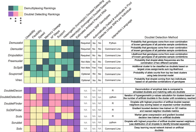
Assessment of each of the demultiplexing and doublet detecting methods. Assessments of a variety of metrics for each of the demultiplexing (top) and doublet detecting (bottom) methods
Framework for improving singlet classifications via method combinations
After identifying the demultiplexing and doublet detecting methods that performed well individually, we next sought to test whether using intersectional combinations of multiple methods would enhance droplet classifications and provide a software platform— Demuxafy —capable of supporting the execution of these intersectional combinations.
We recognise that different experimental designs will be required for each project. As such, we considered this when testing combinations of methods. We considered multiple experiment designs and two different intersectional methods: (1) more than half had to classify a droplet as a singlet to be called a singlet and (2) at least half of the methods had to classify a droplet as a singlet to be called a singlet. Significantly, these two intersectional methods only differ when an even number of methods are being considered. For combinations that include demultiplexing methods, the individual called by the majority of the methods is the individual used for that droplet. When ties occur, the individual is considered ‘unassigned’.
Combining multiple doublet detecting methods improve doublet removal for non-multiplexed experimental designs
For the non-multiplexed experimental design, we considered all possible method combinations (Additional file 1 : Table S12). We identified important differences depending on the number of droplets captured and have provided recommendations accordingly. We identified that DoubletFinder , Scrublet , ScDblFinder and Scds is the ideal combination for balanced droplet calling when less than 2,000 droplets are captured. Scds and ScDblFinder or Scrublet , Scds and ScDblFinder is the best combination when 2,000–10,000 droplets are captured. Scds , Scrublet, ScDblFinder and DoubletDetection is the best combination when 10,000–20,000 droplets are captured and Scrublet , Scds , DoubletDetection and ScDblFinder . It is important to note that even a slight increase in the MCC significantly impacts the number of true singlets and true doublets classified with the degree of benefit highly dependent on the original method performance. The combined method increases the MCC compared to individual doublet detecting methods on average by 0.11 and up to 0.33—a significant improvement in the MCC ( t -test FDR < 0.05 for 95% of comparisons). For all combinations, the intersectional droplet method requires more than half of the methods to consider the droplet a singlet to classify it as a singlet (Fig. 5 ).
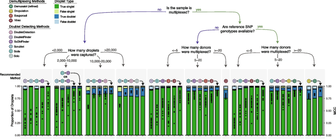
Recommended Method Combinations Dependent on Experimental Design. Method combinations are provided for different experimental designs, including those that are not multiplexed (left) and multiplexed (right), including experiments that have reference SNP genotypes available vs those that do not and finally, multiplexed experiments with different numbers of individuals multiplexed. The each bar represents either a single method (shown with the coloured icon above the bar) or a combination of methods (shown with the addition of the methods and an arrow indicating the bar). The proportion of true singlets, true doublets, false singlets and false doublets for each method or combination of methods is shown with the filled barplot and the MCC is shown with the black points overlaid on the barplot. MCC: Matthew’s Correlation Coefficient
Demuxafy performs better than Chord
Chord is an ensemble machine learning doublet detecting method that uses Scds and DoubletFinder to identify doublets. We compared Demuxafy using Scds and DoubletFinder to Chord and identified that Demuxafy outperformed Chord in pools that contained at least eight donors and was equivalent in pools that contained less than eight donors (Additional file 2 : Fig S14). This is because Chord classifies more droplets as false singlets and false doublets than Demuxafy . In addition, Chord failed to complete for two of the pools that contained 128 multiplexed donors.
Combining multiple demultiplexing and doublet detecting methods improve doublet removal for multiplexed experimental designs
For experiments where 16 or fewer individuals are multiplexed with reference SNP genotypes available, we considered all possible combinations between the demultiplexing and doublet detecting methods except ScDblFinder with known doublets due to its highly similar performance to ScDblFinder (Fig 3 ; Additional file 1 : Table S13). The best combinations are DoubletFinder , Scds , ScDblFinder , Vireo and Demuxalot (refined) (<~5 donors) and Scrublet , ScDblFinder , DoubletDetection , Dropulation and Demuxalot (refined) (Fig. 5 ). These intersectional methods increase the MCC compared to the individual methods ( t -test FDR < 0.05), generally resulting in increased true singlets and doublets compared to the individual methods. The improvement in MCC depends on every single method’s performance but, on average, increases by 0.22 and up to 0.71. For experiments where the reference SNP genotypes are unknown, the individuals multiplexed in the pool with 16 or fewer individuals multiplexed, DoubletFinder , ScDblFinder, Souporcell and Vireo (<~5 donors) and Scds , ScDblFinder , DoubletDetection , Souporcell and Vireo are the ideal methods (Fig. 5 ). These intersectional methods again significantly increase the MCC up to 0.87 compared to any of the individual techniques that could be used for this experimental design ( t -test FDR < 0.05 for 94.2% of comparisons). In both cases, singlets should only be called if more than half of the methods in the combination classify the droplet as a singlet.
Combining multiple demultiplexing methods improves doublet removal for large multiplexed experimental designs
For experiments that multiplex more than 16 individuals, we considered the combinations between all demultiplexing methods (Additional file 1 : Table S14) since only a small proportion of the doublets would be undetectable by demultiplexing methods (droplets that are homogenic; Fig 3 b). To balance doublet removal and maintain true singlets, we recommend the combination of Demuxalot (refined) and Dropulation . These method combinations significantly increase the MCC by, on average, 0.09 compared to all the individual methods ( t -test FDR < 0.05). This substantially increases true singlets and true doublets relative to the individual methods. If reference SNP genotypes are not available for the individuals multiplexed in the pools, Vireo performs the best (≥ 16 multiplexed individuals; Fig. 5 ). This is the only scenario in which executing a single method is advantageous to a combination of methods. This is likely due to the fact that most of the methods perform poorly for larger pool sizes (Fig. 3 c).
These results collectively demonstrate that, regardless of the experimental design, demultiplexing and doublet detecting approaches that intersect multiple methods significantly enhance droplet classification. This is consistent across different pool sizes and will improve singlet annotation.
Demuxafy improves doublet removal and improves usability
To make our intersectional approaches accessible to other researchers, we have developed Demuxafy ( https://demultiplexing-doublet-detecting-docs.readthedocs.io/en/latest/index.html ) - an easy-to-use software platform powered by Singularity. This platform provides the requirements and instructions to execute each demultiplexing and doublet detecting methods. In addition, Demuxafy provides wrapper scripts that simplify method execution and effectively summarise results. We also offer tools that help estimate expected numbers of doublets and provide method combination recommendations based on scRNA-seq pool characteristics. Demuxafy also combines the results from multiple different methods, provides classification combination summaries, and provides final integrated combination classifications based on the intersectional techniques selected by the user. The significant advantages of Demuxafy include a centralised location to execute each of these methods, simplified ways to combine methods with an intersectional approach, and summary tables and figures that enable practical interpretation of multiplexed datasets (Fig. 1 a).
Demultiplexing and doublet detecting methods have made large-scale scRNA-seq experiments achievable. However, many demultiplexing and doublet detecting methods have been developed in the recent past, and it is unclear how their performances compare. Further, the demultiplexing techniques best detect heterogenic doublets while doublet detecting methods identify heterotypic doublets. Therefore, we hypothesised that demultiplexing and doublet detecting methods would be complementary and be more effective at removing doublets than demultiplexing methods alone.
Indeed, we demonstrated the benefit of utilising a combination of demultiplexing and doublet detecting methods. The optimal intersectional combination of methods depends on the experimental design and capture characteristics. Our results suggest super loaded captures—where a high percentage of doublets is expected—will benefit from multiplexing. Further, when many donors are multiplexed (>16), doublet detecting is not required as there are few doublets that are homogenic and heterotypic.
We have provided different method combination recommendations based on the experimental design. This decision is highly dependent on the research question.
Conclusions
Overall, our results provide researchers with important demultiplexing and doublet detecting performance assessments and combinatorial recommendations. Our software platform, Demuxafy ( https://demultiplexing-doublet-detecting-docs.readthedocs.io/en/latest/index.html ), provides a simple implementation of our methods in any research lab around the world, providing cleaner scRNA-seq datasets and enhancing interpretation of results.
PBMC scRNA-seq data
Blood samples were collected and processed as described previously [ 17 ]. Briefly, mononuclear cells were isolated from whole blood samples and stored in liquid nitrogen until thawed for scRNA-seq capture. Equal numbers of cells from 12 to 16 samples were multiplexed per pool and single-cell suspensions were super loaded on a Chromium Single Cell Chip A (10x Genomics) to capture 20,000 droplets per pool. Single-cell libraries were processed per manufacturer instructions and the 10× Genomics Cell Ranger Single Cell Software Suite (v 2.2.0) was used to process the data and map it to GRCh38. Cellbender v0.1.0 was used to identify empty droplets. Almost all droplets reported by Cell Ranger were identified to contain cells by Cellbender (mean: 99.97%). The quality control metrics of each pool are demonstrated in Additional file 2 : Fig S15.
PBMC DNA SNP genotyping
SNP genotype data were prepared as described previously [ 17 ]. Briefly, DNA was extracted from blood with the QIAamp Blood Mini kit and genotyped on the Illumina Infinium Global Screening Array. SNP genotypes were processed with Plink and GCTA before imputing on the Michigan Imputation Server using Eagle v2.3 for phasing and Minimac3 for imputation based on the Haplotype Reference Consortium panel (HRCr1.1). SNP genotypes were then lifted to hg38 and filtered for > 1% minor allele frequency (MAF) and an R 2 > 0.3.
Fibroblast scRNA-seq data
The fibroblast scRNA-seq data has been described previously [ 18 ]. Briefly, human skin punch biopsies from donors over the age of 18 were cultured in DMEM high glucose supplemented with 10% fetal bovine serum (FBS), L-glutamine, 100 U/mL penicillin and 100 μg/mL (Thermo Fisher Scientific, USA).
For scRNA-seq, viable cells were flow sorted and single cell suspensions were loaded onto a 10× Genomics Single Cell 3’ Chip and were processed per 10× instructions and the Cell Ranger Single Cell Software Suite from 10× Genomics was used to process the sequencing data into transcript count tables as previously described [ 18 ]. Cellbender v0.1.0 was used to identify empty droplets. Almost all droplets reported by Cell Ranger were identified to contain cells by Cellbender (mean: 99.65%). The quality control metrics of each pool are demonstrated in Additional file 2 : Fig S16.
Fibroblast DNA SNP genotyping
The DNA SNP genotyping for fibroblast samples has been described previously [ 18 ]. Briefly, DNA from each donor was genotyped on an Infinium HumanCore-24 v1.1 BeadChip (Illumina). GenomeStudioTM V2.0 (Illumina), Plink and GenomeStudio were used to process the SNP genotypes. Eagle V2.3.5 was used to phase the SNPs and it was imputed with the Michigan Imputation server using minimac3 and the 1000 genome phase 3 reference panel as described previously [ 18 ].
Demultiplexing methods
All the demultiplexing methods were built and run from a singularity image.
Demuxalot [ 6 ] is a genotype reference-based single cell demultiplexing method. Demualot v0.2.0 was used in python v3.8.5 to annotate droplets. The likelihoods, posterior probabilities and most likely donor for each droplet were estimated using the Demuxalot Demultiplexer.predict_posteriors function. We also used Demuxalot Demultiplexer.learn_genotypes function to refine the genotypes before estimating the likelihoods, posterior probabilities and likely donor of each droplet with the refined genotypes as well.
The Popscle v0.1-beta suite [ 16 ] for population genomics in single cell data was used for Demuxlet and Freemuxlet demultiplexing methods. The popscle dsc-pileup function was used to create a pileup of variant calls at known genomic locations from aligned sequence reads in each droplet with default arguments.
Demuxlet [ 3 ] is a SNP genotype reference-based single cell demultiplexing method. Demuxlet was run with a genotype error coefficient of 1 and genotype error offset rate of 0.05 and the other default parameters using the popscle demuxlet command from Popscle (v0.1-beta).
Freemuxlet [ 16 ] is a SNP genotype reference-free single cell demultiplexing method. Freemuxlet was run with default parameters including the number of samples included in the pool using the popscle freemuxlet command from Popscle (v0.1-beta).
Dropulation
Dropulation [ 5 ] is a SNP genotype reference-based single cell demultiplexing method that is part of the Drop-seq software. Dropulation from Drop-seq v2.5.1 was implemented for this manuscript. In addition, the method for calling singlets and doublets was provided by the Dropulation developer and implemented in a custom R script available on Github and Zenodo (see “Availability of data and materials”).
ScSplit v1.0.7 [ 7 ] was downloaded from the ScSplit github and the recommended steps for data filtering quality control prior to running ScSplit were followed. Briefly, reads that had read quality lower than 10, were unmapped, were secondary alignments, did not pass filters, were optical PCR duplicates or were duplicate reads were removed. The resulting bam file was then sorted and indexed followed by freebayes to identify single nucleotide variants (SNVs) in the dataset. The resulting SNVs were filtered for quality scores greater than 30 and for variants present in the reference SNP genotype vcf. The resulting filtered bam and vcf files were used as input for the s cSplit count command with default settings to count the number of reference and alternative alleles in each droplet. Next the allele matrices were used to demultiplex the pool and assign cells to different clusters using the scSplit run command including the number of individuals ( -n ) option and all other options set to default. Finally, the individual genotypes were predicted for each cluster using the scSplit genotype command with default parameters.
Souporcell [ 4 ] is a SNP genotype reference-free single cell demultiplexing method. The Souporcell v1.0 singularity image was downloaded via instructions from the gihtub page. The Souporcell pipeline was run using the souporcell_pipeline.py script with default options and the option to include known variant locations ( --common_variants ).
Vireo [ 2 ] is a single cell demultiplexing method that can be used with reference SNP genotypes or without them. For this assessment, Vireo was used with reference SNP genotypes. Per Vireo recommendations, we used model 1 of the cellSNP [ 20 ] version 0.3.2 to make a pileup of SNPs for each droplet with the recommended options using the genotyped reference genotype file as the list of common known SNP and filtered with SNP locations that were covered by at least 20 UMIs and had at least 10% minor allele frequency across all droplets. Vireo version 0.4.2 was then used to demultiplex using reference SNP genotypes and indicating the number of individuals in the pools.
Doublet detecting methods
All doublet detecting methods were built and run from a singularity image.
DoubletDecon
DoubletDecon [ 9 ] is a transcription-based deconvolution method for identifying doublets. DoubletDecon version 1.1.6 analysis was run in R version 3.6.3. SCTransform [ 21 ] from Seurat [ 22 ] version 3.2.2 was used to preprocess the scRNA-seq data and then the Improved_Seurat_Pre_Process function was used to process the SCTransformed scRNA-seq data. Clusters were identified using Seurat function FindClusters with resolution 0.2 and 30 principal components (PCs). Then the Main_Doublet_Decon function was used to deconvolute doublets from singlets for six different rhops—0.6, 0.7, 0.8, 0.9, 1.0 and 1.1. We used a range of rhop values since the doublet annotation by DoubletDecon is dependent on the rhop parameter which is selected by the user. The rhop that resulted in the closest number of doublets to the expected number of doublets was selected on a per-pool basis and used for all subsequent analysis. Expected number of doublets were estimated with the following equation:
where N is the number of droplets captured and D is the number of expected doublets.
DoubletDetection
DoubletDetection [ 14 ] is a transcription-based method for identifying doublets. DoubletDetection version 2.5.2 analysis was run in python version 3.6.8. Droplets without any UMIs were removed before analysis with DoubletDetection . Then the doubletdetection.BoostClassifier function was run with 50 iterations with use_phenograph set to False and standard_scaling set to True. The predicted number of doublets per iteration was visualised across all iterations and any pool that did not converge after 50 iterations, it was run again with increasing numbers of iterations until they reached convergence.
DoubletFinder
DoubletFinder [ 10 ] is a transcription-based doublet detecting method. DoubletFinder version 2.0.3 was implemented in R version 3.6.3. First, droplets that were more than 3 median absolute deviations (mad) away from the median for mitochondrial per cent, ribosomal per cent, number of UMIs or number of genes were removed per developer recommendations. Then the data was normalised with SCTransform followed by cluster identification using FindClusters with resolution 0.3 and 30 principal components (PCs). Then, pKs were selected by the pK that resulted in the largest BC MVN as recommended by DoubletFinder. The pK vs BC MVN relationship was visually inspected for each pool to ensure an effective BC MVN was selected for each pool. Finally, the homotypic doublet proportions were calculated and the number of expected doublets with the highest doublet proportion were classified as doublets per the following equation:
ScDblFinder
ScDblFinder [ 11 ] is a transcription-based method for detecting doublets from scRNA-seq data. ScDblFinder 1.3.25 was implemented in R version 4.0.3. ScDblFinder was implemented with two sets of options. The first included implementation with the expected doublet rate as calculated by:
where N is the number of droplets captured and R is the expected doublet rate. The second condition included the same expected number of doublets and included the doublets that had already been identified by all the demultiplexing methods.
Scds [ 12 ] is a transcription-based doublet detecting method. Scds version 1.1.2 analysis was completed in R version 3.6.3. Scds was implemented with the cxds function and bcds functions with default options followed by the cxds_bcds_hybrid with estNdbl set to TRUE so that doublets will be estimated based on the values from the cxds and bcds functions.
Scrublet [ 13 ] is a transcription-based doublet detecting method for single-cell RNA-seq data. Scrublet was implemented in python version 3.6.3. Scrublet was implemented per developer recommendations with at least 3 counts per droplet, 3 cells expressing a given gene, 30 PCs and a doublet rate based on the following equation:
where N is the number of droplets captured and R is the expected doublet rate. Four different minimum number of variable gene percentiles: 80, 85, 90 and 95. Then, the best variable gene percentile was selected based on the distribution of the simulated doublet scores and the location of the doublet threshold selection. In the case that the selected threshold does not fall between a bimodal distribution, those pools were run again with a manual threshold set.
Solo [ 15 ] is a transcription-based method for detecting doublets in scRNA-seq data. Solo was implemented with default parameters and an expected number of doublets based on the following equation:
where N is the number of droplets captured and D is the number of expected doublets. Solo was additionally implemented in a second run for each pool with the doublets that were identified by all the demultiplexing methods as known doublets to initialize the model.
In silico pool generation
Cells that were identified as singlets by all methods were used to simulate pools. Ten pools containing 2, 4, 8, 16, 32, 64 and 128 individuals were simulated assuming a maximum 20% doublet rate as it is unlikely researchers would use a technology that has a higher doublet rate. The donors for each simulated pool were randomly selected using a custom R script which is available on Github and Zenodo (see ‘Availability of data and materials’). A separate bam for the cell barcodes for each donor was generated using the filterbarcodes function from the sinto package (v0.8.4). Then, the GenerateSyntheticDoublets function provided by the Drop-seq [ 5 ] package was used to simulate new pools containing droplets with known singlets and doublets.
Twenty-one total pools—three pools from each of the different simulated pool sizes (2, 4, 8, 16, 32, 64 and 128 individuals) —were used to simulate different experimental scenarios that may be more challenging for demultiplexing and doublet detecting methods. These include simulating higher ambient RNA, higher mitochondrial percent, decreased read coverage and imbalanced donor proportions as described subsequently.

High ambient RNA simulations
Ambient RNA was simulated by changing the barcodes and UMIs on a random selection of reads for 10, 20 or 50% of the total UMIs. This was executed with a custom R script that is available in Github and Zenodo (see ‘Availability of data and materials’).
High mitochondrial percent simulations
High mitochondrial percent simulations were produced by replacing reads in 5, 10 or 25% of the randomly selected cells with mitochondrial reads. The number of reads to replace was derived from a normal distribution with an average of 30 and a standard deviation of 3. This was executed with a custom R script available in Github and Zenodo (see ‘Availability of data and materials’).
Imbalanced donor simulations
We simulated pools that contained uneven proportions of the donors in the pools to identify if some methods are better at demultiplexing pools containing uneven proportions of each donor in the pool. We simulated pools where 50, 75 or 95% of the pool contained cells from a single donor and the remainder of the pool was even proportions of the remaining donors in the pool. This was executed with a custom R script available in Github and Zenodo (see ‘Availability of data and materials’).
Decrease read coverage simulations
Decreased read coverage of pools was simulated by down-sampling the reads by two-thirds of the original coverage.
Classification annotation
Demultiplexing methods classifications were considered correct if the droplet annotation (singlet or doublet) and the individual annotation was correct. If the droplet type was correct but the individual annotation was incorrect (i.e. classified as a singlet but annotated as the wrong individual), then the droplet was incorrectly classified.
Doublet detecting methods were considered to have correct classifications if the droplet annotation matched the known droplet type.
All downstream analyses were completed in R version 4.0.2.
Availability of data and materials
All data used in this manuscript is publicly available. The PBMC data is available on GEO (Accession: GSE196830) [ 23 ] as originally described in [ 17 ]. The fibroblast data is available on ArrayExpress (Accession Number: E-MTAB-10060) [ 24 ] and as originally described in [ 18 ]. The code used for the analyses in this manuscript are provided on Github ( https://github.com/powellgenomicslab/Demuxafy_manuscript/tree/v4 ) and Zenodo ( https://zenodo.org/records/10813452 ) under an MIT Open Source License [ 25 , 26 ]. Demuxafy is provided as a package with source code available on Github ( https://github.com/drneavin/Demultiplexing_Doublet_Detecting_Docs ) and instructions on ReadTheDocs ( https://demultiplexing-doublet-detecting-docs.readthedocs.io/en/latest/ ) under an MIT Open Source License [ 27 ]. Demuxafy is also available on Zenodo with the link https://zenodo.org/records/10870989 [ 28 ].
Zheng GXY, Terry JM, Belgrader P, Ryvkin P, Bent ZW, Wilson R, et al. Massively parallel digital transcriptional profiling of single cells. Nat Commun. 2017;8:1–12.
Article Google Scholar
Huang Y, McCarthy DJ, Stegle O. Vireo: Bayesian demultiplexing of pooled single-cell RNA-seq data without genotype reference. Genome Biol. 2019;20:273.
Article PubMed PubMed Central Google Scholar
Kang HM, Subramaniam M, Targ S, Nguyen M, Maliskova L, McCarthy E, et al. Multiplexed droplet single-cell RNA-sequencing using natural genetic variation. Nat Biotechnol. 2018;36:89–94.
Article CAS PubMed Google Scholar
Heaton H, Talman AM, Knights A, Imaz M, Gaffney DJ, Durbin R, et al. Souporcell: robust clustering of single-cell RNA-seq data by genotype without reference genotypes. Nat Methods. 2020;17:615–20.
Wells MF, Nemesh J, Ghosh S, Mitchell JM, Salick MR, Mello CJ, et al. Natural variation in gene expression and viral susceptibility revealed by neural progenitor cell villages. Cell Stem Cell. 2023;30:312–332.e13.
Article CAS PubMed PubMed Central Google Scholar
Rogozhnikov A, Ramkumar P, Shah K, Bedi R, Kato S, Escola GS. Demuxalot: scaled up genetic demultiplexing for single-cell sequencing. bioRxiv. 2021;2021.05.22.443646.
Xu J, Falconer C, Nguyen Q, Crawford J, McKinnon BD, Mortlock S, et al. Genotype-free demultiplexing of pooled single-cell RNA-seq. Genome Biol. 2019;20:290.
What is the maximum number of cells that can be profiled?. Available from: https://kb.10xgenomics.com/hc/en-us/articles/360001378811-What-is-the-maximum-number-of-cells-that-can-be-profiled -
DePasquale EAK, Schnell DJ, Van Camp PJ, Valiente-Alandí Í, Blaxall BC, Grimes HL, et al. DoubletDecon: deconvoluting doublets from single-cell RNA-sequencing data. Cell Rep. 2019;29:1718–1727.e8.
McGinnis CS, Murrow LM, Gartner ZJ. DoubletFinder: doublet detection in single-cell RNA sequencing data using artificial nearest neighbors. Cell Syst. 2019;8:329–337.e4.
Germain P-L, Lun A, Meixide CG, Macnair W, Robinson MD. Doublet identification in single-cell sequencing data. 2022;
Bais AS, Kostka D. Scds: Computational annotation of doublets in single-cell RNA sequencing data. Bioinformatics. 2020;36:1150–8.
Wolock SL, Lopez R, Klein AM. Scrublet: computational identification of cell doublets in single-cell transcriptomic data. Cell Syst. 2019;8:281–291.e9.
Shor, Jonathan. DoubletDetection. Available from: https://github.com/JonathanShor/DoubletDetection .
Bernstein NJ, Fong NL, Lam I, Roy MA, Hendrickson DG, Kelley DR. Solo: doublet identification in single-cell RNA-Seq via semi-supervised deep learning. Cell Syst. 2020;11:95–101.e5.
popscle. Available from: https://github.com/statgen/popscle .
Yazar S, Alquicira-Hernandez J, Wing K, Senabouth A, Gordon MG, Andersen S, et al. Single-cell eQTL mapping identifies cell type–specific genetic control of autoimmune disease. Science. 2022;376:eabf3041.
Neavin D, Nguyen Q, Daniszewski MS, Liang HH, Chiu HS, Senabouth A, et al. Single cell eQTL analysis identifies cell type-specific genetic control of gene expression in fibroblasts and reprogrammed induced pluripotent stem cells. Genome Biol. 2021;1–19.
Hao Y, Hao S, Andersen-Nissen E, Mauck WM, Zheng S, Butler A, et al. Integrated analysis of multimodal single-cell data. Cell. 2021;184:3573–3587.e29.
Huang X, Huang Y. Cellsnp-lite: an efficient tool for genotyping single cells. bioRxiv. 2021;2020.12.31.424913.
Hafemeister C, Satija R. Normalization and variance stabilization of single-cell RNA-seq data using regularized negative binomial regression. bioRxiv. 2019;576827.
Stuart T, Butler A, Hoffman P, Hafemeister C, Papalexi E, Mauck WM, et al. Comprehensive integration of single-cell data. Cell. 2019;177:1888–1902.e21.
Powell JE. Single-cell eQTL mapping identifies cell type specific genetic control of autoimmune disease. Datasets. Gene Expression Omnibus. 2022. https://www.ncbi.nlm.nih.gov/geo/query/acc.cgi?acc=GSE196830 .
Nguyen Q, Powell JE. scRNA-seq in 79 fibroblast cell lines and 31 reprogrammed induced pluripotent stem cell lines for sceQTL analysis. Datasets. ArrayExpress. 2021. https://www.ebi.ac.uk/biostudies/arrayexpress/studies/E-MTAB-10060?query=E-MTAB-10060 .
Neavin DR. Demuxafy analyses. Github. 2024. https://github.com/powellgenomicslab/Demuxafy_manuscript/tree/v4 .
Neavin DR. Demuxafy analyses. Zenodo. 2024. https://zenodo.org/records/10813452 .
Neavin D. Demuxafy. Github. 2024. https://github.com/drneavin/Demultiplexing_Doublet_Detecting_Docs .
Neavin D. Demuxafy. Zenodo. 2024. https://zenodo.org/records/10870989 .
McCaughey T, Liang HH, Chen C, Fenwick E, Rees G, Wong RCB, et al. An interactive multimedia approach to improving informed consent for induced pluripotent stem cell research. Cell Stem Cell. 2016;18:307–8.
Download references
Authors’ Twitter handles
Twitter handles: @drneavin (Drew Neavin), @thjimmylee (Jimmy Tsz Hang Lee), @marta_mele_m (Marta Melé)
Peer review information
Wenjing She was the primary editor of this article and managed its editorial process and peer review in collaboration with the rest of the editorial team.
Review history
The review history is available as Additional file 3 .
This work was funded by the National Health and Medical Research Council (NHMRC) Investigator grant (1175781), and funding from the Goodridge foundation. J.E.P is also supported by a fellowship from the Fok Foundation.
Author information
Authors and affiliations.
Garvan-Weizmann Centre for Cellular Genomics, Garvan Institute for Medical Research, Darlinghurst, NSW, Australia
Drew Neavin, Anne Senabouth, Himanshi Arora & Joseph E. Powell
Present address: Statewide Genomics at NSW Health Pathology, Sydney, NSW, Australia
Himanshi Arora
Wellcome Sanger Institute, Wellcome Genome Campus, Hinxton, UK
Jimmy Tsz Hang Lee
Life Sciences Department, Barcelona Supercomputing Center, Barcelona, Catalonia, Spain
Aida Ripoll-Cladellas & Marta Melé
Department of Genetics, University of Groningen, University Medical Center Groningen, Groningen, The Netherlands
Lude Franke
Spatial and Single Cell Systems Domain, Genome Institute of Singapore (GIS), Agency for Science, Technology and Research (A*STAR), Singapore, Republic of Singapore
Shyam Prabhakar
Population and Global Health, Lee Kong Chian School of Medicine, Nanyang Technological University, Singapore, Republic of Singapore
Cancer Science Institute of Singapore, National University of Singapore, Singapore, Republic of Singapore
Bakar Institute for Computational Health Sciences, University of California, San Francisco, CA, USA
Chun Jimmie Ye
Institute for Human Genetics, University of California, San Francisco, San Francisco, CA, USA
Division of Rheumatology, Department of Medicine, University of California, San Francisco, San Francisco, CA, USA
Chan Zuckerberg Biohub, San Francisco, CA, USA
Bioinformatics and Cellular Genomics, St Vincent’s Institute of Medical Research, Fitzroy, Australia
Davis J. McCarthy
Melbourne Integrative Genomics, School of BioSciences–School of Mathematics & Statistics, Faculty of Science, University of Melbourne, Melbourne, Australia
Present address: The Gene Lay Institute of Immunology and Inflammation, Brigham and Women’s Hospital and Harvard Medical School, Boston, MA, USA
Martin Hemberg
UNSW Cellular Genomics Futures Institute, University of New South Wales, Kensington, NSW, Australia
Joseph E. Powell
You can also search for this author in PubMed Google Scholar
sc-eQTLGen Consortium
Contributions.
DRN and JEP conceived the project idea and study design. JTHL, AR, LF, SP, CJY, DJM, MM and MH provided feedback on experimental design. DRN carried out analyses with support on coding from AS. JTHL and AR tested Demuxafy and provided feedback. DRN and JEP wrote the manuscript. All authors reviewed and provided feedback on the manuscript.
Corresponding authors
Correspondence to Drew Neavin or Joseph E. Powell .
Ethics declarations
Ethics approval and consent to participate.
Briefly, all work was approved by the Royal Hobart Hospital, the Hobart Eye Surgeons Clinic, Human Research Ethics Committees of the Royal Victorian Eye and Ear Hospital (11/1031), University of Melbourne (1545394) and University of Tasmania (H0014124) in accordance with the requirements of the National Health & Medical Research Council of Australia (NHMRC) and conformed with the Declaration of Helsinki [ 29 ].
Consent for publication
No personal data for any individual requiring consent for publication was included in this manuscript.
Competing interests
C.J.Y. is founder for and holds equity in DropPrint Genomics (now ImmunAI) and Survey Genomics, a Scientific Advisory Board member for and holds equity in Related Sciences and ImmunAI, a consultant for and holds equity in Maze Therapeutics, and a consultant for TReX Bio, HiBio, ImYoo, and Santa Ana. Additionally, C.J.Y is also newly an Innovation Investigator for the Arc Institute. C.J.Y. has received research support from Chan Zuckerberg Initiative, Chan Zuckerberg Biohub, Genentech, BioLegend, ScaleBio and Illumina.
Additional information
Publisher’s note.
Springer Nature remains neutral with regard to jurisdictional claims in published maps and institutional affiliations.
Supplementary Information
Additional file 1: supplementary tables and legends., additional file 2: supplementary figures and legends., additional file 3..
Review history.
Rights and permissions
Open Access This article is licensed under a Creative Commons Attribution 4.0 International License, which permits use, sharing, adaptation, distribution and reproduction in any medium or format, as long as you give appropriate credit to the original author(s) and the source, provide a link to the Creative Commons licence, and indicate if changes were made. The images or other third party material in this article are included in the article's Creative Commons licence, unless indicated otherwise in a credit line to the material. If material is not included in the article's Creative Commons licence and your intended use is not permitted by statutory regulation or exceeds the permitted use, you will need to obtain permission directly from the copyright holder. To view a copy of this licence, visit http://creativecommons.org/licenses/by/4.0/ . The Creative Commons Public Domain Dedication waiver ( http://creativecommons.org/publicdomain/zero/1.0/ ) applies to the data made available in this article, unless otherwise stated in a credit line to the data.
Reprints and permissions
About this article
Cite this article.
Neavin, D., Senabouth, A., Arora, H. et al. Demuxafy : improvement in droplet assignment by integrating multiple single-cell demultiplexing and doublet detection methods. Genome Biol 25 , 94 (2024). https://doi.org/10.1186/s13059-024-03224-8
Download citation
Received : 07 March 2023
Accepted : 25 March 2024
Published : 15 April 2024
DOI : https://doi.org/10.1186/s13059-024-03224-8
Share this article
Anyone you share the following link with will be able to read this content:
Sorry, a shareable link is not currently available for this article.
Provided by the Springer Nature SharedIt content-sharing initiative
- Single-cell analysis
- Genetic demultiplexing
- Doublet detecting
Genome Biology
ISSN: 1474-760X
- Submission enquiries: [email protected]
- General enquiries: [email protected]
- High School
- You don't have any recent items yet.
- You don't have any courses yet.
- You don't have any books yet.
- You don't have any Studylists yet.
- Information
Group assignment - Fin 555- to analyze the future potential of particular stock
Introduction to technical analysis (fin 555), universiti teknologi mara.
Students also viewed
- Group Assignment FIN555
- TEST 1 FIN555 JULY2023 - tes1
- FIN 552 Trading GAME ( Group Assignment)
- Afifah Elias 2018 861588
- [FIN555] Individual Assignment
- FIN555 _ Individual Assignment_TECHNICAL ANALYSIS_Individual assignment
Related documents
- 4C Siti Nurul Asyikin Binti Hassan 2020993671 A1
- Group Assignment
- MOCK Trading Report - individual assestments
- Nestle FULL FIN555 - graft and chart
- Group Shafinaz Assignment 3 RBA2424A
- Assignment 1.docx - Google Docs
Related Studylists
Preview text.
UNIVERSITI TEKNOLOGI MARA (UiTM) CAWANGAN MELAKA KAMPUS BANDARAYA MELAKA
SEMESTER 1 SESI 2020/
Oktober 2020 – februari 2021, introduction to technical analysis, assignment 3:, group assignment, prepared by:, muhammad danial bin ahmad fadzil 2019871442, nur syahirah binti rahamad 2019489076, ahmad hijaz bin amir firdaus 2019660066, suhaidah binti sulaiman 2019461798, muhammad firdaus bin osman 2018250946, muhammad hisyam bin amiruddin 2019848522, submitted to:, amirudin bin mohd nor, trading plan & spot trading opportunities, padini (7052), sideways to downtrend: is there opportunity still, drbhicom (1619).
Analysis 2 Based on the chart above, we can observe the performance of this stock where the bullish run ended in late September 2020 where the Bollinger band begins to narrow. Then, in October 2020, prices begin to drop, however, there is a bullish signal as we detected there is a positive divergence where prices drop while stochastic has higher lows. Then, on the 9th of November 2020, opportunity has shown itself as the MACD line crossed over the MACD signal line coinciding with the MACD histogram convergence above the 0-line. This was confirmed on the oversold position below the 20% level in the stochastic indicator. Then, on 14the December 2020, the bull run ends as the price crossed above the upper Bollinger band coinciding with the MACD line crossing below the MACD signal line marking a sell signal. So, what is next for this stock DRBHICOM (1619)? As in terms of buying the stock, it is not the time yet to buy the stock because currently, it is trading at RM1 which is below the RM1 support 1 line, which is quite a significant support line. Volume of shares traded has been declining while the price is declining. Other indicators such as MACD and Stochastic oscillator is showing that it is not time yet to buy. The MACD line has not cross over the MACD signal line and the divergence of the MACD histogram is pretty tall below the 0-line which indicates the downtrend is still strong. The stochastic indicator is still below the 20% line which is an oversold position but not a point of entry yet as it has not cross above the 20% line. The stochastic indicator is also supported by the RSI indicator which agrees that this stock is oversold below 30% level (RSI). However, the stock is not ripe to be bought as of now because both the indicators have not established a point of entry. Even the ROC says the momentum of this stock is low and since it is below the 0-line, it is on a downtrend. The stock is on a strong downtrend as it broke away from its sideways position and goes on a downtrend, it is the beginning of a short-term bearish signal as the Bollinger band continues to widen. The strategy is to buy this stock once MACD and stochastic coincide with each other and take profit at resistance level RM2. The stock however is traded below the first support level, we will then observe if the momentum carries on below the second support line at RM1, whether there will be a reversal or not. If there is a reversal, then we can buy this stock, you can check out the stochastic and RSI indicators whether they have crossed above their oversold positions or check out the MACD line whether the MACD line crosses above the MACD signal line. The rating for this stock is a SELL and the stop loss level is at the first support level at RM1.
Analysis 3 The performance of CSCSTEL (5094) is impressive, but before that, from the late July to early November 2020, it has been on a sideways trend with not much activity, this is confirmed by a narrow Bollinger band. Then, the price per share grew from RM0 to RM1, which is a huge increase from early November to mid-December 2020. As the Bollinger band widens, the MACD line is above the MACD signal line and the ROC indicates and uptrend with high momentum. Then on the 4th of January 2021, the MACD line crosses below the MACD signal line with the convergence below the 0- line. This coincides with the overbought position of the Bollinger band and the low momentum on the ROC indicator. The RSI indicator also shows the momentum of this stock being very good where it signals to buy the stock at entry on the 9th of November 2020. Then, you can observe there is a negative divergence, which is stating that it begins to become a bearish signal. This bearish divergence materialized as the RSI peaks decline over time while the price of the stock increases. That continues on later to become a downtrend, around this time, the overbought position was reached on 28th December 2020 and from there, the bear caught up and went on a downtrend. The stochastic indicator is in most occasions coincide with the RSI indicator, it is very much the same position for overbought and oversold. The RSI indicator is in an overbought position above the 70% line since 9th November 2020 to 21st December 2020. The stochastic indicator also is at the overbought position above 80% around that month. This tells us that both of these momentum indicators say that this stock is gaining good momentum, and the entry price is at RM0 according to both of these indicators. However, the exit timing is different for both RSI and stochastic. Our trading plan is to hold this stock, because, we believe that there will be a reversal going to occur. Although the price is currently traded on RM1 which is above support line of RM1, and, is traded on the lower Bollinger band range. We believe that this stock will bounce back in the near future. However, be cautious when you execute a trade like this, that is why we are not recommending this stock as a buy, but it is still technically solid to not turn it to a sell. If you are holding this stock, do not sell just yet. But, do not buy at this range, because you may make a loss. By far, this is a neutral stance, hoping that this stock will have a reversal. Stop-loss is at RM1 or below and take profit is at RM1 which is the resistance level. We hope to see the ROC indicator will show higher momentum to back the trade up and crosses the 0-line again to indicate an uptrend. The rate for this stock is a HOLD for these reasons.
OPPORTUNITY WILL PREVAIL
The volume game: scalping for profit, dsonic (5216).
Analysis 5 The MA crossover of this share indicates a sell on the 17th of August 2020 where the stock is traded at RM0. This is when DSONIC (5216) is going on a downtrend which began on 17th August until 28th September 2020, a really sharp decline in price from RM0 to RM0. Meanwhile on another indicator which is the MACD indicator, it recommended a buy at RM0 on 1st October 2020 where the MACD line crosses above the MACD signal line and MACD histogram is converging from below to above the 0-line. The volume of stocks traded however seems to be the major influencer for the price movement of this stock. It seems that every time the stock goes up in price, the volume goes up, if the stock goes down, there seems to be a drop in volume of shares traded. The stochastic and RSI indicators for this stock only reflects the momentum of this stock’s performance. It does not give good entry or exit positions for this stock. The performance of this stock in particular is not really good for the past 6 months where it is bearish in the months of July to early September 2020. Then it is sideways from September 2020 to January 2021. Not much activity except signs of short-term trading where the price is heavily influenced by volume of shares traded. This stock also exposes that there are limits to the momentum indicators as well as moving averages. The momentum indicators are at the end of the day, lagging indicators. You can see that a lot of the indicators are too little too late to show the buy and sell signals. There is one good sell signal in January 2021 which is accurate where there was an MA crossover short-term crosses below long-term moving average but, the entry or buy signals are off, if you buy it early, you will still make a loss. Our buy signal according to stochastic is on the 28th of December 2020 at RM0. Sell signal or exit price is also at RM0, by theory, there is no profit, but taking into account with brokerage fee, you would lose money. Even MACD in January 2021 is not really clear whether it is a buy or a sell because the MACD line is very close to the MACD signal line, although MACD gave a good buy position on 1st October 2020, it did not give good sell positions. This stock is good for short-term trading as only volumes influence this stock. Even the oversold position for stochastic below the 20% line or oversold RSI below the 30% line is a bad indicator. Currently, the price of this stock is traded at RM0, strategy is intraday or scalping following volumes traded daily to gain profits from small price change. This strategy is risky and not recommended to beginner traders. This stock is also very risky and is not recommended to trade for beginners. The rate for this stock is a SELL.
Analysis 6 This stock had a great performance for the past 6 months where it has been bullish. The bull run is where there have been new higher highs from July 2020 to January 2021. The MA Envelope is been on an uptrend, the ROC indicator shows that most of the time, the price is on an uptrend because the ROC line is most of the time above the 0-line with high momentums supporting the ROC line. The RSI indicates that it has reached overbought level above 70% twice, which this stock is good to trade with. There is a little bit of a slowdown in between the months of late October to mid- December 2020, where there has been a negative divergence occurring between the RSI indicator together with the price chart. The price goes up while the peaks of RSI moved downwards. But then, momentum was gained back again and the price reaches its second overbought position above the 70% line. On the 10th of August 2020, when the MA Envelope upper moving average was breached, showing that the stock is overbought, it is confirmed by the MACD indicator. As you can see that the MACD line crossed below the MACD signal line which is a sell signal. This is coincided with the MACD histogram where a convergence from above the 0-line converges towards the 0-line eventually breaking below the 0-line. On the 11th of January 2020 however, it states that the MACD line has crossed above the MACD signal line which is a buy signal. Although this stock generally has been on an uptrend for 6 months, the stock does have 2 support lines. The first support line is at RM2, it will not reach that level any time soon if the stock keeps having higher highs. While the second support line is at RM2 which is a significant support line. Only in bad circumstances, that support line will be breached. This stock would be a good hold if the price of stock is not overbought. Even the volume is pretty high and is not showing signs of slowing down. Currently the stock is trading at RM3 as of 26th January 2021, which is an overbought signal because it is above the upper moving average which is confirmed by all of the indicators, even ROC says it has a high momentum and a strong uptrend. The recommended strategy is to sell the stock in a few more days, because it is already overbought, and confirmed by MA Envelope with RSI and stochastic indicator. But, be aware if there are sudden reversals for this stock which may be bad as it might have the momentum to face a loss, so, a stop loss at RM2, the first support line. The rate for this stock is a SELL, however, even if the rate is a sell, it is best to hold on for a little while more and prepare for a sell is because most indicators are not close to exit price.
Course : Introduction to technical Analysis (FIN 555)
University : universiti teknologi mara, this is a preview.
Access to all documents
Get Unlimited Downloads
Improve your grades
Get 30 days of free Premium
Share your documents to unlock

Why is this page out of focus?

IMAGES
VIDEO
COMMENTS
FIN555 _ Individual Assignment_TECHNICAL ANALYSIS_Individual assignment. Introduction to technical Analysis 100% (6) 10. ... 0 FIN555 2 Format Assignment 1 Chap 11-16 2022 Full Time; Fin 552 pyq exercise; TEST2 JAN2023 - test; FIN555 CIMB ASSIGNMENT; Related documents. Stock Performance OF Sapura Energy Berhad;
INDIVIDUAL ASSIGNMENT 1 PREPARED BY: NAME : FATIN AQILAH QISTINA BINTI ABD HALIM MATRIC NO : 2019256958 GROUP : BA242 4A PREPARED FOR: PUAN SUHAILY MAIZAN ABDUL MANAF. TABLE OF CONTENT. Analysis of IOI Corp Bhd 3-Analysis of Westport Holdings Bhd 1- CONTENT PAGE; Analysis of IOI Corp Bhd 3-Analysis of IHH Healthcare Bhd 5-Analysis of Kobay ...
FIN555 Individual Assignment Nasha Shafridah 2021112481 BA2424B; FIN555 Individual Assignment; FIN555 Assignment 2; Sample of Individual Assignment 1 Report; Assignment 2; FIN 555 Assignment 1 - Chart Patterns ; Preview text. FACULTY OF BUSINESS AND MANAGEMENT UNIVERSITI TEKNOLOGI MARA KAMPUS DUNGUN
FIN555 Individual Assignment .pdf. Universiti Teknologi Mara. FIN 555. FIN420 individual.pdf. Universiti Teknologi Mara. FIN 420. View More. BBA (HONS) FINANCE: SEMESTER SEPTEMBER 2019 - JANUARY 2020 FUNDAMENTAL OF TECHNICAL ANALYSIS (FIN555) Stock Technical Analysis: AMMB Holdings Berhad Nashima Aman Nashima binti Aman, 2018683198, UiTM ...
individual assignment fin555 mohamad zaid bin ismail.pdf. introduction of technical analysis (fin555) individual assignment company analysis prepared by: mohamad zaid bin ismail 2020993625 prepared for: sir anuar bin wahab class: kba2424a date of submission: 22nd july 2021 1 table of contents 1. introduction. 3
View FIN555 Assignment 1 (SHARIFAH ATHIRAH).docx from FIN 555 at Universiti Teknologi Mara. FIN555 ASSIGNMENT 1 "SUPPORT & RESISTANCE, TRENDLINES & PRICE PATTERN" PREPARED BY: NAME : SHARIFAH ATHIRAH. AI Homework Help. Expert Help. Study Resources. ... FIN555 Individual Assignment .pdf. Universiti Teknologi Mara. FIN 555. ASSIGNMENT 3 BA242 4D ...
INDIVIDUAL ASSIGNMENT FIN555 - Free download as PDF File (.pdf), Text File (.txt) or read online for free. This document is an individual assignment analyzing the technical indicators of MR D.I.Y. Group (M) Berhad stock from January to December 2021. It identifies buy and sell signals using four indicators: moving average, Bollinger bands, relative strength index with rate of change, and ...
ASSIGNMENT 1 FIN555 (AutoRecovered) - Free download as Word Doc (.doc / .docx), PDF File (.pdf), Text File (.txt) or read online for free. FIN555
FIN555 _ Individual Assignment_TECHNICAL ANALYSIS_Individual assignment. Mandatory assignments 100% (6) 7. MOCK Trading Report - individual assestments. Mandatory assignments 90% (10) 11. FIN555 individual Assignment. Tutorial work 100% (2) 6. exercise in technical analysis. Tutorial work 100% (1) 8.
About Press Copyright Contact us Creators Advertise Developers Terms Privacy Press Copyright Contact us Creators Advertise Developers Terms Privacy
1 bachelor of business administration (hons) finance faculty of business and management fin555 introduction of technical analysis individual assignment prepared by: name student id 1) muhammad naim bin mohd nahar 2020608754 2) muhammad khaizuran bin abdul halil 2021114751 3) nur amira adilah binti hasni 2021131971 group: ba2424c prepared for: dr.
FIN555 - Free download as Word Doc (.doc / .docx), PDF File (.pdf), Text File (.txt) or read online for free. FIN555
(fin555) individual assignment 1 group: d2ba2424d prepared by: nurhaziera najwa binti mohd zaki student number: 2022787137 prepared for: dr. md khairu amin bin ismail submission date: 20 may 2023 table of contents introduction.
2 INTRODUCTION TO THE STOCK 1.1 Company Summary GDEX Bhd, formerly GD Express Carrier Bhd, is a Malaysia-based investment holding company. Nature of the business is providing express carrier and logistic services. GDEX Bhd was listed on the Bursa Malaysia Securities Berhad in 13 May 2005 and their stock code is GDEX (0078). The company's segments are Express delivery, Logistics and Property ...
technical analysis individual assignment; FIN555 Assignment 1; Envelopes AND Bollinger Bands; Group Assignment FIN555; TEST 1 FIN555 JULY2023 - tes1; FIN 552 Trading GAME ( Group Assignment) Related documents. Afifah Elias 2018 861588; 4C Siti Nurul Asyikin Binti Hassan 2020993671 A1;
FIN555 ASSIGNMENT GROUP 5 - Free download as PDF File (.pdf), Text File (.txt) or read online for free.
1 1.0 ACKNOWLEDGMENT First of all, I would like to say thank you to my beloved lecturer, Madam Nurulhaida Binti Johan who teaches me in subject Introduction to Technical Analysis, code subject FIN555. She gives us information about this subject and giving us some guidelines for doing this individual assignment. I am really grateful because I have completed this individual assignment within the ...
Recent innovations in single-cell RNA-sequencing (scRNA-seq) provide the technology to investigate biological questions at cellular resolution. Pooling cells from multiple individuals has become a common strategy, and droplets can subsequently be assigned to a specific individual by leveraging their inherent genetic differences. An implicit challenge with scRNA-seq is the occurrence of ...
0 FIN555 2 Format Assignment 1 Chap 11-16 2022 Full Time; Fin 552 pyq exercise; TEST2 JAN2023 - test; Related documents. ... exercise in technical analysis; FIN555 individual Assignment; INTRODUCTION TO TECHNICAL ANALYSIS WHICH MEANS, CHAPTER 1 OF THE FIN552 UITM MERBOK KEDAH; Preview text. TUTORIAL CHAPTER 1: ANSWER ALL QUESTIONS. i. Draw the ...
Individual Assignment FIN552 - Free download as Word Doc (.doc / .docx), PDF File (.pdf), Text File (.txt) or read online for free. Scribd is the world's largest social reading and publishing site.
INDIVIDUAL ASSIGNMENT 1 & 2 INSTRUCTION: Choose ONE public listed company. Do the following: Assignment 1 (20%): Perform fundamental analysis on the chosen company.Your contents must include: 1.1.Company stock summary 1.2.Business Review 1.3.Financial Performance Review 1.4.Stock Prices and Dividend Review 1.5.Analyst stock recommendation based on fundamental analysis TAKE NOTE: Follow the ...
IMAN FIRDAUS MOHD KHAIROL ANUAR_INDIVIDUAL ASSIGNMENT FIN555.docx. Therrell School Of Health And Science. FIN 552. View More. INTRODUCTION TO TECHNICAL ANALYSIS (FIN 555) ... FINS5542 Assignment 1 Kevin Xi.docx. University of New South Wales. FINS 5542. FINS5542 Assignment 1 Kevin Xi.docx.
FIN555 _ Individual Assignment_TECHNICAL ANALYSIS_Individual assignment; Related documents. 4C Siti Nurul Asyikin Binti Hassan 2020993671 A1; Group Assignment; MOCK Trading Report - individual assestments; Nestle FULL FIN555 - graft and chart; Group Shafinaz Assignment 3 RBA2424A; Assignment 1.docx - Google Docs;
FIN555 Individual Assignment .pdf. Universiti Teknologi Mara. FIN 555. MBA 550 FINAL PROJECT MODULE 9-1 CHELSEA DIGGS.docx. Solutions Available. Southern New Hampshire University. MBA 550. Trading Strategy - Vladimir Poltoratskiy.pdf. College of Lake County. CMM MISC.